. 2021 Sep 8;22(18):9699.
doi: 10.3390/ijms22189699.

And Yet It Moves: Oxidation of the Nuclear Autoantigen La/SS-B Is the Driving Force for Nucleo-Cytoplasmic Shuttling

Affiliations

And Yet It Moves: Oxidation of the Nuclear Autoantigen La/SS-B Is the Driving Force for Nucleo-Cytoplasmic Shuttling

Nicole Berndt et al. Int J Mol Sci. .

Abstract

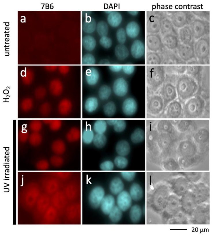
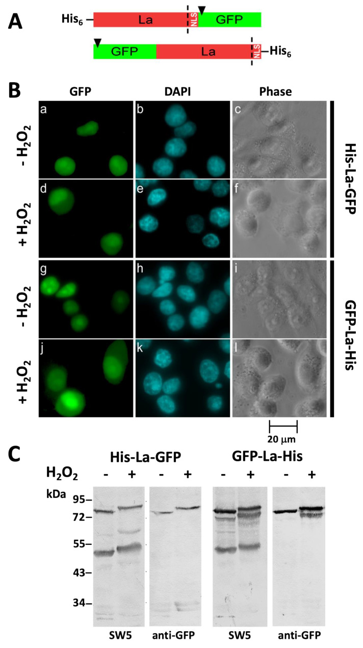
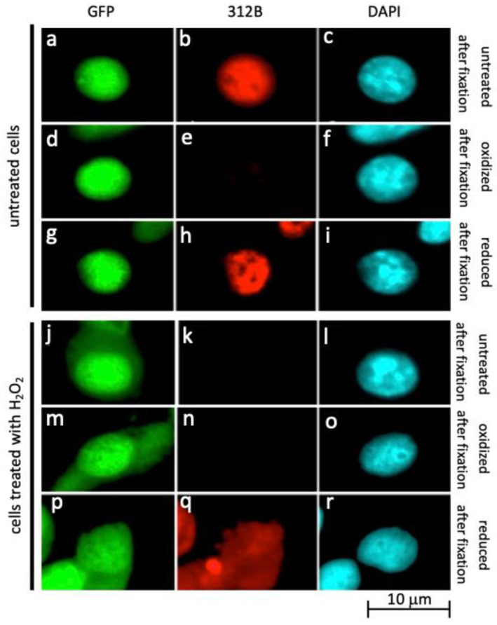
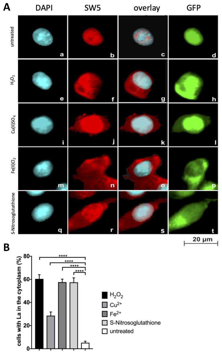
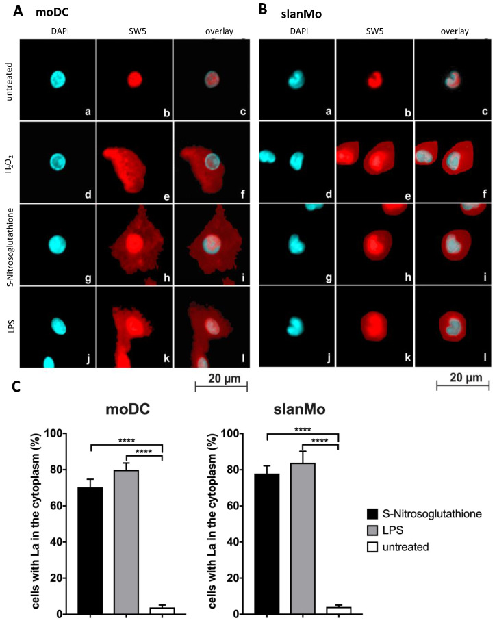
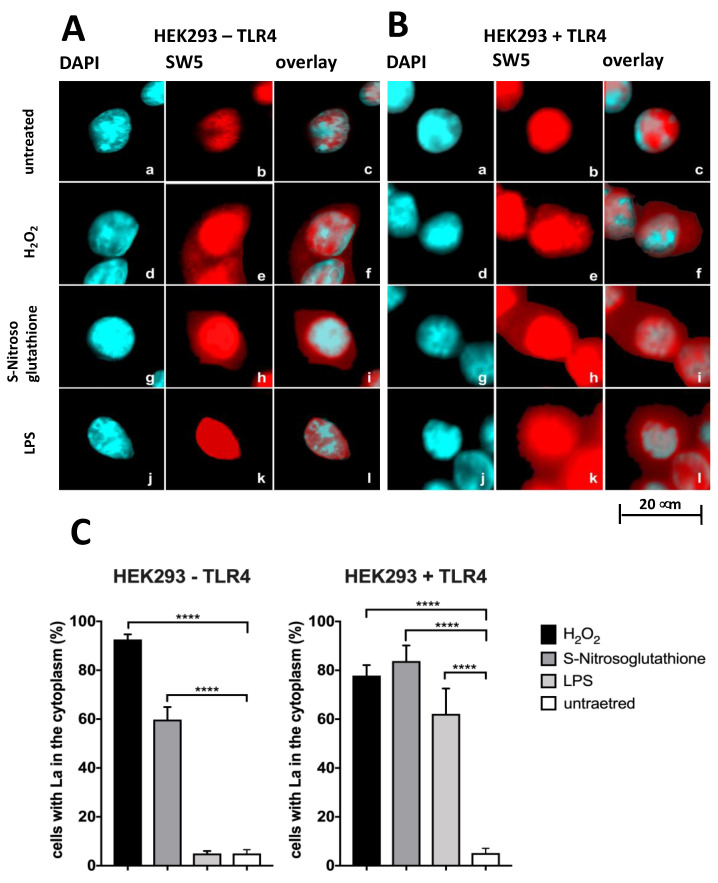
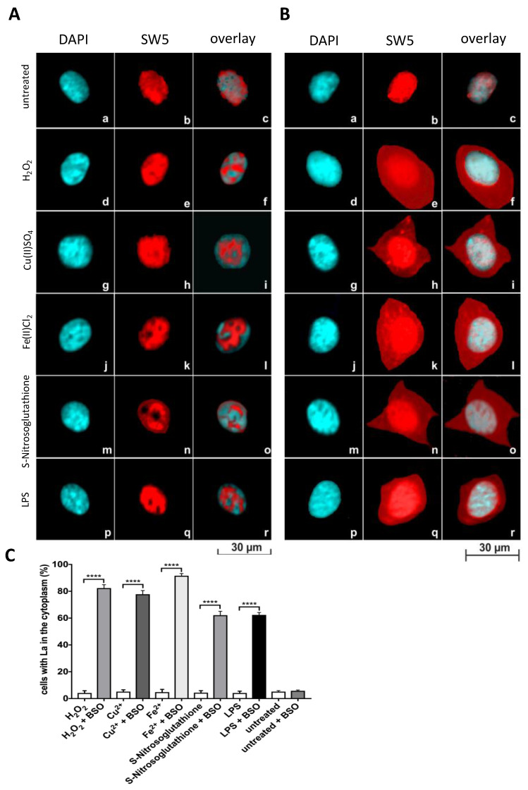
Decades ago, we and many other groups showed a nucleo-cytoplasmic translocation of La protein in cultured cells. This shuttling of La protein was seen after UV irradiation, virus infections, hydrogen peroxide exposure and the Fenton reaction based on iron or copper ions. All of these conditions are somehow related to oxidative stress. Unfortunately, these harsh conditions could also cause an artificial release of La protein. Even until today, the shuttling and the cytoplasmic function of La/SS-B is controversially discussed. Moreover, the driving mechanism for the shuttling of La protein remains unclear. Recently, we showed that La protein undergoes redox-dependent conformational changes. Moreover, we developed anti-La monoclonal antibodies (anti-La mAbs), which are specific for either the reduced form of La protein or the oxidized form. Using these tools, here we show that redox-dependent conformational changes are the driving force for the shuttling of La protein. Moreover, we show that translocation of La protein to the cytoplasm can be triggered in a ligand/receptor-dependent manner under physiological conditions. We show that ligands of toll-like receptors lead to a redox-dependent shuttling of La protein. The shuttling of La protein depends on the redox status of the respective cell type. Endothelial cells are usually resistant to the shuttling of La protein, while dendritic cells are highly sensitive. However, the deprivation of intracellular reducing agents in endothelial cells makes endothelial cells sensitive to a redox-dependent shuttling of La protein.

Keywords: La/SS-B autoantigen; anti-La/SS-B antibodies; autoimmunity; primary Sjögren’s Syndrome; redox sensor; shuttling of La protein; systemic lupus erythematosus.


Conflict of interest statement

All authors declare no conflicts of interest.

Figures

Figure 1
Figure 1
Translocation of La protein to the cytoplasm after UV irradiation. Fixed cells were stained with (a) the anti-La mAb 7B6 or (b) DAPI. (c) Phase-contrast image. (d) Prior to the staining with the anti-La mAb 7B6 La protein was oxidized by washing the fixed cells with PBS containing H2O2. (e) DAPI staining of fixed cells. (f) Phase-contrast image. (g) Cells were UV irradiated. Nine min after irradiation, the cells were fixed and stained with the anti-La mAb 7B6. (h) DAPI staining of fixed cells. (i) phase-contrast image. (j) Cells were UV irradiated. Two h after irradiation, the cells were fixed and stained with the anti-La mAb 7B6. (k) DAPI staining of fixed cells. (l) Phase contrast image.
Figure 2
Figure 2
La protein is not cleaved under oxidative conditions. (A) La protein contains potential proteolytic cleavage sites (dashed line) upstream of the nuclear location signal (NLS). Two different GFP–La-His fusion proteins were constructed in which GFP is fused either upstream or downstream of the La reading frame. In case GFP is fused to the N-terminus of La protein, a His-Tag is fused to the C-terminus of La protein resulting in the fusion protein GFP–La-His. In case GFP is fused to the C-terminus of La protein a His-Tag is fused to the N-terminus of La protein resulting in the fusion protein His-La-GFP. (B) Human HeLa cells expressing either the His-La-GFP fusion construct (af) or the GFP–La-His fusion construct (gl). Cells were either untreated (ac; gi) or treated with H2O2 (df; jl). (C) SDS-PAGE and immunoblotting of total extracts from cells expressing either the His-La-GFP fusion construct (His-La-GFP) or the GFP–La-His fusion construct (GFP-La-His). Total extracts were blotted against either the anti-La mAb SW5 (SW5) or an anti-GFP Ab (anti-GFP). Extracts were obtained from transfected cells which were either untreated (−) or treated with H2O2 (+) prior to the preparation of the extracts.
Figure 3
Figure 3
La protein is oxidized in the cytoplasm. (aq) IF microscopy of mouse 3T3 cells overexpressing His-La-GFP (a,d,g,j,m,p) stained with the anti-La mAb 312B (b,e,h,k,n,q) or DAPI (c,f,i,l,o,r). (ac) Untreated fixed cells. (df) Untreated cells oxidized with H2O2 after fixation. (gi) Untreated cells oxidized with H2O2 after fixation followed by a reduction with ß-mercaptoethanol. (jl) Oxidized living cells, untreated after fixation. (mo) Oxidized living cells, oxidized after fixation. (pr) Oxidized living cells, reduced after fixation.
Figure 4
Figure 4
Translocation of La protein to the cytoplasm and its dependence on oxidative conditions. (A) Immunofluorescence analysis. (ad). Untreated mouse 3T3 cells overexpressing His-La-GFP. (eh) Cells treated with H2O2 or (il) CuSO4, or (mp) FeCl2, or (qt) S-Nitrosoglutathione. Fixed cells were stained with (a,e,i,m,q) DAPI or (b,f,j,n,r) the anti-La mAb SW5. Overlay of DAPI and SW5 staining (c,g,k,o,s) as well as GFP fluorescence (d,h,l,p,t). (B) Quantitative evaluation of the percentage of cells showing cytoplasmic staining comparing untreated cells with cells after the respective treatment. p Value < 0.0001 = ****.
Figure 5
Figure 5
Shuttling of La protein in moDCs or slan monocytes (slanMos). (A,B) MoDCs and slanMos were stained with DAPI (a,d,g,j) or the anti-La mAb SW5 (b,e,h,k). The overlay of both channels is shown (c,f,i,l). (ac) Untreated cells. (df) Cells treated with H2O2. (gi) Cells treated with S-Nitrosoglutathione. (jl) Cells treated with LPS. (C) Quantitative evaluation. p Value < 0.0001 = ****.
Figure 6
Figure 6
Shuttling of La protein in dependence on Toll-like Receptor/Ligand ligation. (A,B) HEK293T cells lacking (HEK293T-TLR4) or overexpressing (HEK293T+TLR4) TLR4 were stained with DAPI (a,d,g,j) or the anti-La mAb SW5 (b,e,h,k). Overlay of both is shown (c,f,i,l). (ac) Untreated cells. (df) Cells treated with H2O2. (gi) Cells treated with S-Nitrosoglutathione. (jl) Cells treated with LPS. (C) Quantitative evaluation. p Value < 0.0001 = ****.
Figure 7
Figure 7
Shuttling of La protein in human umbilical vein endothelial cells (HUVEC). (A,B) HUVECs grown in the absence (A) or presence (B) of buthionine-sulfoximine (BSO). HUVECs were stained with DAPI (a,d,g,j,m,p) or the anti-La mAb SW5 (b,e,h,k,n,q). Overlay of both channels is shown in addition (c,f,i,l,o,r). (ac) Untreated cells. (df) Cells treated with H2O2. (gi) Cells treated with Cu(II)SO4. (jl) Cells treated with Fe(II)Cl2. (mo) Cells treated with S-Nitrosoglutathione. (jl) Cells treated with LPS. (C) Quantitative evaluation. p Value < 0.0001 = ****.

References

    1. Jones B.R. Lacrimal and salivary precipitating antibodies in Sjogren’s syndrome. Lancet. 1958;11:773–776. doi: 10.1016/S0140-6736(58)91480-6. - DOI - PubMed
    1. Anderson J.R., Gray K.G., Beck J.S., Kinnear W.F. Precipitating autoantibodies in Sjogren’s syndrome. Lancet. 1961;2:456–460. doi: 10.1016/S0140-6736(61)92430-8. - DOI - PubMed
    1. Anderson J.R., Gray K.G., Beck J.S., Buchanan W.W., McElhinney A.J. Precipitating autoantibodies in the connective tissue diseases. Ann. Rheum. Dis. 1962;21:360–369. doi: 10.1136/ard.21.4.360. - DOI - PMC - PubMed
    1. Clark G., Reichlin M., Tomasi T.B. Characterization of a soluble cytoplasmic antigen reactive with sera from patients with systemic lupus erythematosus. J. Immunol. 1969;102:117–122. - PubMed
    1. Mattioli M., Reichlin M. Heterogeneity of RNA protein antigens reactive with sera of patients with systemic lupus erythematosus: Description of a cytoplasmic nonribosomal antigen. Arthritis Rheum. 1974;17:421–429. doi: 10.1002/art.1780170413. - DOI - PubMed

MeSH terms

LinkOut - more resources