. 2021 Sep 10;22(18):9811.
doi: 10.3390/ijms22189811.

The Cdc14 Phosphatase Controls Resolution of Recombination Intermediates and Crossover Formation during Meiosis

Affiliations

The Cdc14 Phosphatase Controls Resolution of Recombination Intermediates and Crossover Formation during Meiosis

Paula Alonso-Ramos et al. Int J Mol Sci. .

Abstract

Meiotic defects derived from incorrect DNA repair during gametogenesis can lead to mutations, aneuploidies and infertility. The coordinated resolution of meiotic recombination intermediates is required for crossover formation, ultimately necessary for the accurate completion of both rounds of chromosome segregation. Numerous master kinases orchestrate the correct assembly and activity of the repair machinery. Although much less is known, the reversal of phosphorylation events in meiosis must also be key to coordinate the timing and functionality of repair enzymes. Cdc14 is a crucial phosphatase required for the dephosphorylation of multiple CDK1 targets in many eukaryotes. Mutations that inactivate this phosphatase lead to meiotic failure, but until now it was unknown if Cdc14 plays a direct role in meiotic recombination. Here, we show that the elimination of Cdc14 leads to severe defects in the processing and resolution of recombination intermediates, causing a drastic depletion in crossovers when other repair pathways are compromised. We also show that Cdc14 is required for the correct activity and localization of the Holliday Junction resolvase Yen1/GEN1. We reveal that Cdc14 regulates Yen1 activity from meiosis I onwards, and this function is essential for crossover resolution in the absence of other repair pathways. We also demonstrate that Cdc14 and Yen1 are required to safeguard sister chromatid segregation during the second meiotic division, a late action that is independent of the earlier role in crossover formation. Thus, this work uncovers previously undescribed functions of the evolutionary conserved Cdc14 phosphatase in the regulation of meiotic recombination.

Keywords: CDK1; Cdc14; Cdc20; Cdc5; Holliday junction; Mus81; Ndt80; Sgs1; Yen1; aneuploidy; meiotic recombination.


Conflict of interest statement

The authors declare no conflict of interest. The funders had no role in the design of the study; in the collection, analyses, or interpretation of data; in the writing of the manuscript, or in the decision to publish the results.

Figures
Figure 1
Figure 1
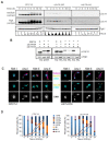
cdc14-HA behaves as a specific sporulation-deficient separation-of-function allele of CDC14. (A) The cdc14-HA allele does not perturb normal growth conditions when cultivated at 25 °C, 30 °C and 37 °C in rich media (YPD). (B) Homozygous cdc14-HA diploids do not form asci containing spores under standard conditions for sporulation. Scale bar represents 1 µm. (C) Percentage of sporulation in CDC14 (JCY840), cdc14-HA (JCY844) or cdc14-md (JCY2376). Error bars represent the standard deviation of the mean (SEM) calculated from at least three independent experiments. A minimun of 300 cells per strain were counted. (D) cdc14-HA diploid cells divide mitotically but lack di-tyrosine autofluorescence induced by UV light after several days of incubation in SPM media at 30 °C. (E) FACS analysis of DNA content shows that cdc14-HA cells complete meiotic DNA replication with identical kinetics as CDC14 diploid cells. (F) cdc14-HA cells undergo meiotic nuclear divisions at subtly slower kinetics, and reduced frequencies, than CDC14 cells. Error bars represent the SEM over the mean values plotted. (G) Di-tyrosine autofluorescence of different mutant combinations as well as the control strains grown and sporulated on plates at 25 °C. cdc14-1 homozygous diploids (JCY2353) sporulate at high efficiency under semi-permissive temperature forming preferentially tetrads whereas cdc14-HA homozygous diploids (JCY844) do not sporulate. Combinations, and variable copy number, of the mutant genes can rescue the sporulation defect to different degrees (JCY2365/JCY2354/JCY2356).
Figure 2
Figure 2
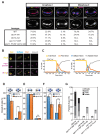
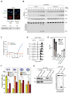
Diminished Cdc14 protein levels in cdc14-HA meiotic cells compromise SPB integrity following meiotic divisions. (A) Dynamics of Cdc14 protein levels throughout meiosis in synchronous time courses for CDC14 (JCY2232), cdc14-HA (JCY2231), and cdc14-md (JCY2389). Higher contrast was applied to visualize residual protein levels for all three strains. Immunodetection of Pgk1 was used as a loading control. FACS histograms for each time-point are depicted at the bottom to show the degree of synchrony reached in each culture. (B) Variability of protein levels in mitotic versus meiotic cells in different CDC14 alleles. Exponentially growing cells show similar Cdc14 levels, excluding cdc14-HA, which has lower overall protein levels (left panel). Direct comparison between the three alleles CDC14 (JCY2232); cdc14-HA (JCY2231) and cdc14-md (JCY2389) in exponentially growing mitotic cells (Exp.) and meiotic cultures immediately after they were transferred to SPM (Mei0). The loading control for each lane was determined using Pgk1. (C) Visualization of Spc29-CFP at SPBs, tubulin, and DNA, in wild-type (JCY892) and cdc14-HA (JCY893) cells fixed at different stages of meiosis at 30 °C. Scale bar represents 1 µm. (D) cdc14-HA cells develop meiosis I and II spindles with near wild-type kinetics. Lack of spore formation in cdc14-HA meiotic cells parallels the loss of SPB’s structural integrity.
Figure 3
Figure 3
Mis-segregation of sister chromatids at MII in meiosis-deficient cdc14 mutant cells. (A(iiii)) Frequency of cells presenting connected DAPI masses at anaphase I in wild type (JCY902), cdc14-HA (JCY904), cdc14-md (PRY132), spo11-Y135F (PRY53) and spo11-Y135F cdc14-HA (PRY55). (A(iv,v)) Frequency of cells presenting connected nuclei at metaphase II. Meiotic-deficient cdc14-HA mutants (GGY54) present higher frequencies of unresolved nuclear divisions at late anaphase I and at metaphase II than wild type (GGY53). (B) Representative images of distinct morphological patterns for Rec8-GFP in meiosis. Full: bright nuclear localization. Diffuse: faint staining within the stretched nucleus. Peri: Rec8-GFP only visible at peri-centromeric locations. Absent: no Rec8-GFP signal. Pds1-tdTomato and CNM67-tdTomato were used to follow Pds1 accumulation/degradation and SPB number/location. (C) Temporal distribution of the distinct morphological categories of Rec8-GFP shown in (B) for CDC14 (JCY2406; n = 57 cells) and cdc14-HA (JCY2404; n = 52 cells). (D) Unequal distribution of homozygous chromosome II-linked GFP markers at lys2 locus in cdc14-HA mutant cells (JCY2330) denotes increased chromosome missegregation in comparison with wild-type cells (JCY2331). (E) Similar distribution of homozygous CENIV-linked GFP markers at trp1 locus in meiosis-deficient cdc14-HA mutant cells (JCY2286) in comparison with wild-type cells (JCY2284) denotes correct homolog disjunction in anaphase I. (F) Unequal distribution of heterozygous CENIV-GFP marker in meiosis-deficient cdc14-HA tetranucleated mutant cells (JCY2327) denotes increased sister chromatid missegregation in comparison with wild-type cells (JCY2326). Statistical significance of differences was calculated by two-tailed t-test, assuming unequal variances (* p < 0.05; ** p < 0.01; *** p < 0.001; **** p < 0.0001; n.s.: not significant). (G) Sporulation defects in cdc14-HA (JCY844) or cdc14-md (JCY2376) can be partly alleviated by eliminating recombination (JCY2270/JCY2280/PRY151). CDC14 (JCY840). Error bars represent SEM over the calculated mean value from three independent experiments. A minimum of 300 cells per strain were counted.
Figure 4
Figure 4
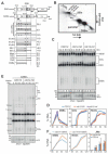
Analysis of meiotic recombination in meiotic-deficient cdc14 mutants. (A) Schematic illustration of recombination analysis and intermediates at the HIS4LEU2 hotspot (see Results for more details). (B) Schematic representation of DNA species commonly observed after analyzing the HIS4LEU2 hotspot by 2D-gel electrophoresis. ((C); top) Representative 1D-gel Southern blot for analysis of DSBs and COs at the HIS4LEU2 hotspot using XhoI as the restriction site. Wild-type strain (JCY2232), cdc14-HA (JCY2231), and cdc14-md (JCY2389). ((C); bottom) Analysis of COs and NCOs at the HIS4LEU2 hotspot using XhoI and NgoMVI in the same strains as in (C). (D) Quantification of DSBs, COs and NCOs from the Southern blots shown in (C). (E) Representative 1D-gel Southern blot image for analysis of DSBs, COs (top) and NCOs (bottom) at the HIS4LEU2 hotspot in CDC14 ndt80Δ strain (JCY2390) and cdc14-HA ndt80Δ (JCY2385) strains. Asterisk indicates meiosis-specific non-characterized recombination products. (F) Quantification of DSBs, NCOs and JMs from the Southern blots shown in (E). Error bars represent SEM over the calculated mean value.
Figure 5
Figure 5
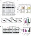
Analysis of recombination intermediates in cdc14 mutants shows early and late accumulation of JMs. (A) Representative Southern blots depicting 1D-gels at the HIS4LEU2 hotspot following induction of CDC5 in prophase-arrested ndt80Δ mus81Δ cdc14-HA cells (JCY2440) leads to inefficient CO and NCO formation compared to ndt80Δ mus81Δ (JCY2442). (B) Quantification of COs and NCOs from Southern blots shown in (A). (C) Representative Southern blots depicting 2D-gels at the HIS4LEU2 hotspot in sgs1-md mms4Δ (JCY2444), sgs1-md mms4Δ cdc14-md (JCY2446), sgs1-md mms4Δ yen1Δ (JCY2448) and sgs1-md mms4Δ cdc14-md YEN1ON (JCY2529) 24 h into meiosis. Arrows mark IH-dHJs and lines mark mc-JMs (see Figure 4A,B). (D) Quantification of total JMs in the strains shown in (C) from at least two independent gels. Error bars represent SEM over the calculated mean value. (E) Representative 1D-gel Southern blot images for analysis of crossovers at the HIS4LEU2 hotspot for all strains shown in (C) and for cdc14-md (JCY2389). (F) Quantification of COs from at least three different image acquisitions such as that depicted in (E). Error bars represent SEM over the calculated mean value.
Figure 6
Figure 6
The absence of Cdc14 prevents the activation of Yen1 during meiotic divisions. (A) Quantification of distinct localization patterns of Yen1 at meiosis I. Scale bar represents 1 µm. (B) Analysis of expression levels and nuclease activity of Yen1 in CDC14 (YML7692) and cdc14-HA (YML7693) meiotic cells. Soluble extracts were prepared from YEN1-Myc9 strains at 2 h intervals after transfer into sporulation medium (SPM). Following anti-Myc immunoaffinity purification (IP), the IPs were analyzed by Western blotting and tested for nuclease activity using Cy3-labeled Holliday junction DNA as a substrate. Upper panel: Western blots of the cell extracts, with detection of Yen1-myc9, Cdc5, and Pgk1 (loading control). Lower panel: HJ resolution assay. The experiment shown is representative of two independent experiments. (C) Percentage of HJ cleavage in Yen1 IPs during meiosis in wild type and cdc14-HA. (D) Evolution of DNA content during meiosis from strains in (B). (E) Unrestrained resolution of recombination intermediates by Yen1ON improves sporulation in cdc14-HA (JCY2164) and cdc14-md (PRY182) cells. Frequency of asci containing one, two, three and four spores in the strains of the indicated genotypes. Error bars represent SEM over the calculated mean value from three independent experiments. A minimum of 300 cells per strain were counted. (F) Frequency of cells presenting connected DAPI masses at anaphase I (left panel) or sister chromatid mis-segregation (right panel) in YEN1ON (PRY123/PRY99), cdc14-HA (JCY2327/PRY55) and cdc14-HA YEN1ON (PRY121/PRY108). Statistical significance of differences was calculated by two-tailed t-test, assuming unequal variances (* p < 0.05; ** p < 0.01; *** p < 0.001; **** p < 0.0001; n.s.: not significant). (G) Poor HJ resolution in cdc14-HA (YML7693) is efficiently restored by the presence of Yen1ON (JCY2421). Western blot analysis of Yen1/Yen1ON immunoprecipitates is shown on the left panels. HJ resolution assay is shown on the right panel. Quantification of resolution efficiency is displayed at the bottom. Resolution data arise from two independent experiments. Asterisks in (B) and (G) indicate labelled DNA strands.
Figure 7
Figure 7
Analysis of recombination intermediates in the cdc20-md mutant reveals an early meiotic role for Cdc14 and Yen1. (A) Representative Southern blots depicting 2D-gels at the HIS4LEU2 hotspot in CDC14 cdc20-md sgs1-md (JCY2480), cdc20-md sgs1-md yen1 (JCY2469), cdc20-md sgs1-md cdc14-md (JCY2508), cdc20-md sgs1-md mms4 (JCY2480), cdc20-md sgs1-md mms4 yen1 (JCY2478) and cdc20-md sgs1-md mms4 cdc14-md (JCY2502). Arrows mark IH-dHJs and lines mark mc-JMs (see Figure 4A,B). (B) Quantification of total JMs in the strains shown in (A) from several independent gels. Error bars display SEM over the mean values plotted. (C) Representative 1D-gel Southern blot images for analysis of crossovers at the HIS4LEU2 hotspot for all strains shown in (A). (D) Quantification of COs from at least three different image acquisitions, such as that depicted in (C). Error bars represent SEM over the calculated mean value.
Figure 8
Figure 8
Model for CDC14-dependent resolution of recombination intermediates via multiple mechanisms. Contribution of Cdc14 to the correct disjunction of recombining chromosomes during meiosis. Early in prophase I, chromosomes initiate homologous recombination. 3′-resected ssDNA overhangs invade an intact template with the help of recombinases. The displacement of the intact strand from the template allows for the formation of D-loops, which can be stabilized, allowing for DNA synthesis. Next, stabilized branched DNA molecules might be disrupted by the action of the anti-recombinases. Reannealing of the extended 3′-ssDNA overhang with the resected complementary strand, followed by further DNA synthesis will lead to the repair and ligation of the broken DNA duplex, giving rise to NCOs via SDSA. ZMM stabilization of JMs is followed by the resolution of dHJs through the MutLγ, class I, dedicated CO pathway. Ndt80-dependent expression, and activation, of Cdc5 triggers MutLγ resolvase activity. Unresolved linkages between bivalents that persist until metaphase/anaphase I are mostly resolved by the action of the SSEs, Mus81-Mms4. Slx1-Slx4, Top3-Rmi1 as well as Cdc14/Yen1 also contribute to the correct resolution of chromosomal entanglements between homologs during MI. Residual chromatid intertwining between sister chromatids during the second meiotic division will be removed by the action of Cdc14/Yen1. Gradual implementation of Yen1 activity during both divisions by Cdc14 will transfer Yen1 inactive population to its active/nuclear-enriched form.

References

    1. Keeney S., Giroux C.N., Kleckner N. Meiosis-specific DNA double-strand breaks are catalyzed by Spo11, a member of a widely conserved protein family. Cell. 1997;88:375–384. doi: 10.1016/S0092-8674(00)81876-0. - DOI - PubMed
    1. Bishop D.K. Multiple mechanisms of meiotic recombination. Cell. 2006;127:1095–1097. doi: 10.1016/j.cell.2006.11.029. - DOI - PubMed
    1. Zakharyevich K., Ma Y., Tang S., Hwang P.Y., Boiteux S., Hunter N. Temporally and biochemically distinct activities of Exo1 during meiosis: Double-strand break resection and resolution of double Holliday junctions. Mol. Cell. 2010;40:1001–1015. doi: 10.1016/j.molcel.2010.11.032. - DOI - PMC - PubMed
    1. Zakharyevich K., Tang S., Ma Y., Hunter N. Delineation of joint molecule resolution pathways in meiosis identifies a crossover-specific resolvase. Cell. 2012;149:334–347. doi: 10.1016/j.cell.2012.03.023. - DOI - PMC - PubMed
    1. Sanchez A., Adam C., Rauh F., Duroc Y., Ranjha L., Lombard B., Mu X., Wintrebert M., Loew D., Guarne A., et al. Exo1 recruits Cdc5 polo kinase to MutLgamma to ensure efficient meiotic crossover formation. Proc. Natl. Acad. Sci. USA. 2020;117:30577–30588. doi: 10.1073/pnas.2013012117. - DOI - PMC - PubMed

MeSH terms