Review
. 2021 Sep 10;22(18):9807.
doi: 10.3390/ijms22189807.

Targeting Autophagy with Natural Products as a Potential Therapeutic Approach for Cancer

Affiliations
Review

Targeting Autophagy with Natural Products as a Potential Therapeutic Approach for Cancer

Md Abdul Alim Al-Bari et al. Int J Mol Sci. .

Abstract

Macro-autophagy (autophagy) is a highly conserved eukaryotic intracellular process of self-digestion caused by lysosomes on demand, which is upregulated as a survival strategy upon exposure to various stressors, such as metabolic insults, cytotoxic drugs, and alcohol abuse. Paradoxically, autophagy dysfunction also contributes to cancer and aging. It is well known that regulating autophagy by targeting specific regulatory molecules in its machinery can modulate multiple disease processes. Therefore, autophagy represents a significant pharmacological target for drug development and therapeutic interventions in various diseases, including cancers. According to the framework of autophagy, the suppression or induction of autophagy can exert therapeutic properties through the promotion of cell death or cell survival, which are the two main events targeted by cancer therapies. Remarkably, natural products have attracted attention in the anticancer drug discovery field, because they are biologically friendly and have potential therapeutic effects. In this review, we summarize the up-to-date knowledge regarding natural products that can modulate autophagy in various cancers. These findings will provide a new position to exploit more natural compounds as potential novel anticancer drugs and will lead to a better understanding of molecular pathways by targeting the various autophagy stages of upcoming cancer therapeutics.

Keywords: anticancer drugs; autophagy; autophagy modulators; mTOR signaling; natural products; resveratrol; ω-3 PUFAs.


Conflict of interest statement

The authors declare no conflict of interest.

Figures

Figure 1
Figure 1
Molecular mechanisms of various stages of autophagy. Autophagy is activated in response to various cellular stresses and is triggered by a decrease in rapamycin complex 1 (mTORC1) activity due to the activation of AMP-activated protein kinase (AMPK) or p53 signaling. mTORC1 suppresses the activity of Unc-51-like autophagy activating kinase 1 (ULK1) complex. Therefore, inhibition of mTORC1 causes the initialization of the ULK1-mediated formation of the isolation (autophagosomal) membrane (IM) in association with the class III phosphatidylinositide 3-kinase (PI3K) complex (PI3KC3). The IM expands into an autophagosome (AP) with a double-layer membrane, which can engulf any cellular component, including proteins, damaged organelles and lipid droplets. The AP merges with the lysosome (via LAMP-1, 2), forming autophagolysosome (APL) or autolysosome (AL), and resulting in the degradation of the cargo by cathepsins and the autophagic lysosome reformation (ALR). The nucleation, elongation and maturation of the IM are dependent on two ubiquitin-like conjugation systems (ATG12 and ATG8), which involve multiple autophagy proteins, including Beclin1, ATG5, ATG16 and MT-associated protein 1 light chain 3 (LC3). The AL provides an acidic milieu for hydrolytic enzymes to digest the engulfed components. Nuclear localization of transcription factor EB (TFEB) is critical to the formation of lysosomes and to the enhanced expression of autophagy proteins. Importantly, autophagy could be selective of mitochondria (mitophagy) or ER (ER-phagy) [7]. However, the detailed mechanisms of this selected autophagy are beyond the scope of this study.
Figure 2
Figure 2
Ultrastructural features of upregulated autophagy in ethanol-treated Sertoli cells (ETR SCs). TEM of the control (A) and ETRs (BG). The histogram (H) shows a significant increase in the number of autophagic vacuoles (AVs) in ETR SCs. The long black arrows indicate autophagosomes with a double limiting membrane (arrow heads) (magnified in the inset in (C)). The short arrows indicate autolysosomes. The broken arrows in (E) show multilamellar bodies, while the white arrows in (G) show autophagosomes containing fragmented mitochondria. Note the characteristic perinuclear localization of AVs. S: SC nucleus; L: lysosome; M: mitochondria; LD: lipid droplet. AVs include autophagosomes and autolysosomes. * p < 0.05. This was reprinted from Reference [3].
Figure 3
Figure 3
Expression of LC3 in stromal cells of serious human ovarian carcinoma using IHC. The black arrows indicate LC3-II puncta in stromal cells, whereas the red arrows mark ovarian cancer cells. The framed area is magnified in the inset. BV, blood vessel. The avidin biotin complex (ABC) IHC method is performed. The diaminobenzidine (DAB) is used as a chromogen (brown reaction), whereas the hematoxylin (blue) is applied for nuclear counterstaining.
Figure 4
Figure 4
Elevated expression and nuclear translocation of TFEB in ETR SCs. (A) The IF of TFEB expressions in the control (a) and ETRs (b). The insets present higher magnifications of the framed areas. Note the overexpression of TFEB (white arrows) in the SC nuclei of ETRs. (B) Immunogold labeling of TFEB (black arrows, 15 nm gold particles) in the control (c) and ETR SCs (d). (C) Western blot of TFEB in the control and ETR tests (n = 3). (D) Histogram showing a significant increase in TFEB expression in the ETR tests. * p < 0.05 (t-test). (E) IHC showing TFEB nuclear translocation in ETR SCs (part of a seminiferous tubule), confirming the IF and IEM results (A,B). S: SC nucleus; L: Leydig cell. This was reprinted from Reference [43].

References

    1. Pohl C., Dikic I. Cellular quality control by the ubiquitin-proteasome system and autophagy. Science. 2019;366:818–822. doi: 10.1126/science.aax3769. - DOI - PubMed
    1. Saftig P., Klumperman J. Lysosome biogenesis and lysosomal membrane proteins: Trafficking meets function. Nat. Rev. Mol. Cell Biol. 2009;10:623–635. doi: 10.1038/nrm2745. - DOI - PubMed
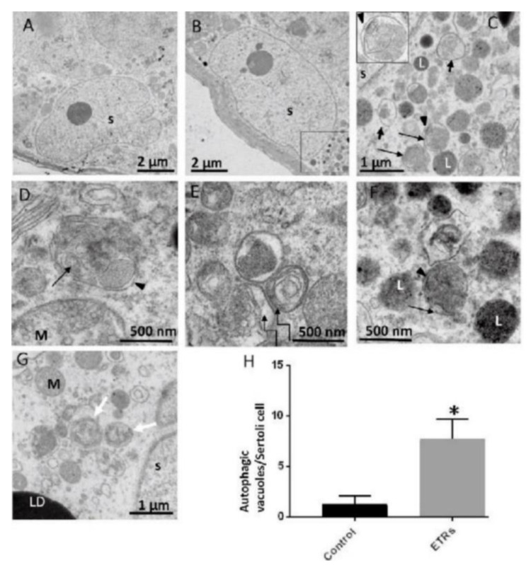
    1. Horibe A., Eid N., Ito Y., Hamaoka H., Tanaka Y., Kondo Y. Upregulated autophagy in Sertoli cells of ethanol-treated rats is associated with induction of inducible nitric oxide synthase (iNOS), androgen receptor suppression and germ cell apoptosis. Int. J. Mol. Sci. 2017;18:1061. doi: 10.3390/ijms18051061. - DOI - PMC - PubMed
    1. Levy J.M.M., Towers C.G., Thorburn A. Targeting autophagy in cancer. Nat. Rev. Cancer. 2017;17:528–542. doi: 10.1038/nrc.2017.53. - DOI - PMC - PubMed
    1. Kaur J., Debnath J. Autophagy at the crossroads of catabolism and anabolism. Nat. Rev. Mol. Cell Biol. 2015;16:461–472. doi: 10.1038/nrm4024. - DOI - PubMed