Review
. 2021 Sep 14;22(18):9904.
doi: 10.3390/ijms22189904.

Tryptophan: From Diet to Cardiovascular Diseases

Affiliations
Review

Tryptophan: From Diet to Cardiovascular Diseases

Nada Joe Melhem et al. Int J Mol Sci. .

Abstract

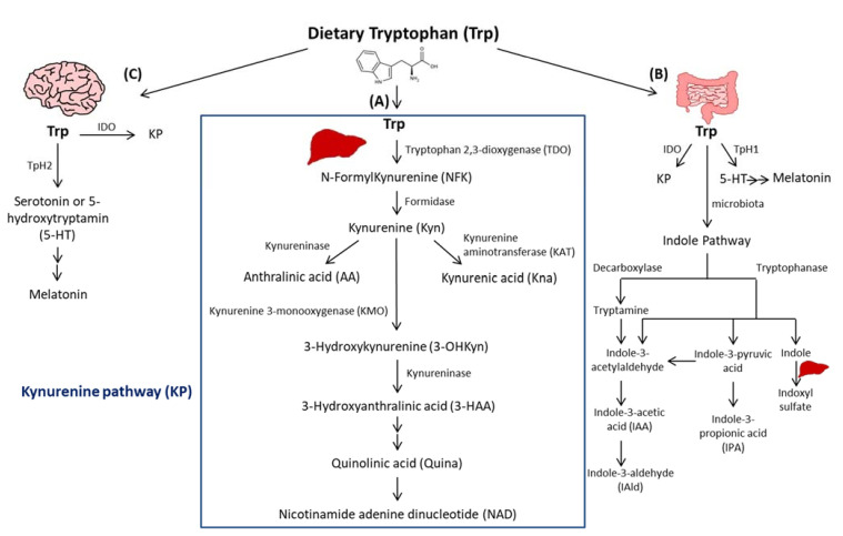
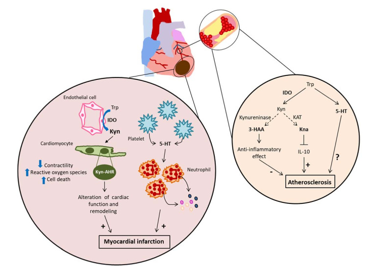
Cardiovascular disease (CVD) is one of the major causes of mortality worldwide. Inflammation is the underlying common mechanism involved in CVD. It has been recently related to amino acid metabolism, which acts as a critical regulator of innate and adaptive immune responses. Among different metabolites that have emerged as important regulators of immune and inflammatory responses, tryptophan (Trp) metabolites have been shown to play a pivotal role in CVD. Here, we provide an overview of the fundamental aspects of Trp metabolism and the interplay between the dysregulation of the main actors involved in Trp metabolism such as indoleamine 2, 3-dioxygenase 1 (IDO) and CVD, including atherosclerosis and myocardial infarction. IDO has a prominent and complex role. Its activity, impacting on several biological pathways, complicates our understanding of its function, particularly in CVD, where it is still under debate. The discrepancy of the observed IDO effects could be potentially explained by its specific cell and tissue contribution, encouraging further investigations regarding the role of this enzyme. Thus, improving our understanding of the function of Trp as well as its derived metabolites will help to move one step closer towards tailored therapies aiming to treat CVD.

Keywords: IDO; atherosclerosis; cardiometabolic diseases; cardiovascular disease; kynurenine; myocardial infarction; tryptophan; tryptophan catabolism.


Conflict of interest statement

The authors declare no conflict of interest.

Figures

Figure 1
Figure 1
Simplified illustration of the tryptophan degradation in different peripheral organs. Tryptophan (Trp) is predominantly converted into kynurenine (Kyn) pathway (KP) by Tryptophan 2, 3-dioxygenase (TDO) in the liver (A) and by indoleamine 2, 3-dioxygenase 1 (IDO) in extrahepatic organs. In the gastrointestinal tract, a small amount of Trp is converted by gut microbiota through the action of the enzymes tryptophanase and decaboxylase, into indole and its derivatives and into tryptamine (B). Indole metabolites could be converted in the liver into indoxyl sulfate. Another fraction of Trp is converted through Trp hydroxylase 1 (TpH 1) into serotonin in the gut (B) and through Tph2 in the brain (C) then to melatonin.
Figure 2
Figure 2
Potential mechanism of actions of Trp metabolites in CVD. In ischemia-induced myocardial infarction (MI), induction of IDO in endothelial cells is responsible of paracrine effects on cardiomyocyte apoptosis through an aryl hydrocarbon receptor (AHR)-dependent manner, as well as an alteration of cardiomyocyte contractility. This impacts on cardiac remodeling and function. In MI, serotonin (5-HT) produced by platelets activates neutrophil degranulation, which aggravates MI. In atherosclerosis, Kyn-derived metabolites, 3-HAA (hydroxyanthranilic acid) has been shown to exert protective effect, whereas another Trp-derived metabolite, kynurenic acid (Kna) has been shown to have deleterious effects on plaque development through the down-regulation of the anti-inflammatory cytokine, IL-10.

References

    1. Roth G.A., Johnson C., Abajobir A., Abd-Allah F., Abera S.F., Abyu G., Ahmed M., Aksut B., Alam T., Alam K., et al. Global, Regional, and National Burden of Cardiovascular Diseases for 10 Causes, 1990 to 2015. J. Am. Coll. Cardiol. 2017;70:1–25. doi: 10.1016/j.jacc.2017.04.052. - DOI - PMC - PubMed
    1. Theodorou K., Boon R.A. Endothelial Cell Metabolism in Atherosclerosis. Front. Cell Dev. Biol. 2018;6:82. doi: 10.3389/fcell.2018.00082. - DOI - PMC - PubMed
    1. Thygesen K., Alpert J.S., Jaffe A.S., Chaitman B.R., Bax J.J., Morrow D.A., White H.D. Fourth Universal Definition of Myocardial Infarction (2018) J. Am. Coll. Cardiol. 2018;72:2231–2264. doi: 10.1016/j.jacc.2018.08.1038. - DOI - PubMed
    1. Ibanez B., James S., Agewall S., Antunes M.J., Bucciarelli-Ducci C., Bueno H., Caforio A.L.P., Crea F., Goudevenos J.A., Halvorsen S., et al. 2017 ESC Guidelines for the management of acute myocardial infarction in patients presenting with ST-segment elevation: The Task Force for the management of acute myocardial infarction in patients presenting with ST-segment elevation of the European Society of Cardiology (ESC) Eur. Heart J. 2018;39:119–177. doi: 10.1093/eurheartj/ehx393. - DOI - PubMed
    1. Ross R. Atherosclerosis—An Inflammatory Disease. N. Engl. J. Med. 1999;340:115–126. doi: 10.1056/nejm199901143400207. - DOI - PubMed

Substances