. 2021 Sep 15;22(18):9951.
doi: 10.3390/ijms22189951.

Downregulated TNF-α Levels after Cryo-Thermal Therapy Drive Tregs Fragility to Promote Long-Term Antitumor Immunity

Affiliations

Downregulated TNF-α Levels after Cryo-Thermal Therapy Drive Tregs Fragility to Promote Long-Term Antitumor Immunity

Yue Lou et al. Int J Mol Sci. .

Abstract

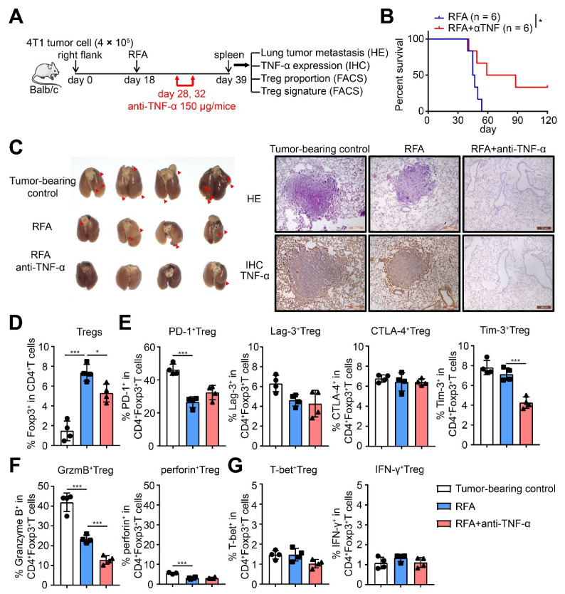
Immunotherapy has emerged as a therapeutic pillar in tumor treatment, but only a minority of patients get benefit. Overcoming the limitations of immunosuppressive environment is effective for immunotherapy. Moreover, host T cell activation and longevity within tumor are required for the long-term efficacy. In our previous study, a novel cryo-thermal therapy was developed to improve long-term survival in B16F10 melanoma and s.q. 4T1 breast cancer mouse models. We determined that cryo-thermal therapy induced Th1-dominant CD4+ T cell differentiation and the downregulation of Tregs in B16F10 model, contributing to tumor-specific and long-lasting immune protection. However, whether cryo-thermal therapy can affect the differentiation and function of T cells in a s.q. 4T1 model remains unknown. In this study, we also found that cryo-thermal therapy induced Th1-dominant differentiation of CD4+ T cells and the downregulation of effector Tregs. In particular, cryo-thermal therapy drove the fragility of Tregs and impaired their function. Furthermore, we discovered the downregulated level of serum tumor necrosis factor-α at the late stage after cryo-thermal therapy which played an important role in driving Treg fragility. Our findings revealed that cryo-thermal therapy could reprogram the suppressive environment and induce strong and durable antitumor immunity, which facilitate the development of combination strategies in immunotherapy.

Keywords: TNF-α; Treg fragility; cryo-thermal therapy.


Conflict of interest statement

The authors declare no conflict of interest.

Figures

Figure 1
Figure 1
Cryo-thermal therapy induced long-term antitumor protection in 4T1-bearing mice. (A) Kaplan–Meier survival curve of tumor-bearing control, traditional RFA or cryo-thermal therapy treated mice. Approximately 4 × 10⁵ 4T1 cells were injected subcutaneously into the right flank of female Balb/c mouse. Then, 18 days later, mice were randomly allocated into three groups, and treated by RFA or cryo-thermal therapy, and untreated mice were as control. Kaplan–Meier survival curve was compared using log-rank tests. * p < 0.05, ** p < 0.01. n = 6 for each group. (B) H&E staining of the lung of tumor-bearing control (39 days after inoculation), RFA or cryo-thermal treated group (21 days after treatment).
Figure 2
Figure 2
Cryo-thermal therapy promoted CD4+ T cell-mediated antitumor immune response. (AC) The percentages of CD4+ T cells of splenic lymphocytes (B) and the proliferation (C) of CD4+ T cells were detected by flow cytometry on day 21 after cryo-thermal therapy or RFA treatment. For CD4+ T cells proliferation assay, splenic CD4+ T cells were isolated from tumor-bearing control (39 days after inoculation), RFA or cryo-thermal (21 days after treatment) treated mice by using microbeads. The CD4+ T cells were labeled with CFSE and then cultured with αCD3 (1 ng/mL) for 24 h. The proliferation of CD4+ T cells (CFSE) was detected by flow cytometry. (DI) The percentages of CD4 Th1 (D), CTL (E), Th17 (F), Th2 (G), Tregs (H), and Tfh (I) subsets in spleen were detected by flow cytometry. * p < 0.05, ** p < 0.01, *** p < 0.001, **** p < 0.0001. n = 4 for each group.
Figure 3
Figure 3
Cryo-thermal therapy drove the fragile phenotype of Tregs and down-regulated its suppressive function. (AD) The expression level of inhibitory checkpoint molecules (A), suppressive cytokines Granzyme B and perforin (B), and T-bet and IFN-γ expression (C) on splenic Tregs from tumor-bearing control (39 days after inoculation), RFA or cryo-thermal therapy (21 days after treatment) were detected by flow cytometry. (D) Splenic Tregs from tumor-bearing control (39 days after inoculation), RFA or cryo-thermal therapy (21 days after treatment) and splenic CD8+ T cells from naïve mice were isolated through microbeads. CD8+ T cells were labeled with CFSE and then co-cultured with Tregs in different E/T ratio. αCD3 (1 ng/mL) was used to stimulated the proliferation of CD8+ T cells. After 72 h, the proliferated CD8+ T cells (CFSE-) were tested by flow cytometry. * p < 0.05, ** p < 0.01, *** p < 0.001, **** p < 0.0001 compared with the E/T ratio of 0:1. &&& p < 0.001, &&&& p < 0.0001 compared with the same E/T ratio of group RFA. N = 4 for each group.
Figure 4
Figure 4
The level of serum TNF-α was decreased after cryo-thermal therapy. (A) The level of serum TNF-α from tumor-bearing control, RFA or cryo-thermal treated mice on different time points was detected by western blot (up), and the gray statistics of three independent experiments (down). (B) The level of serum TNF-α from tumor-bearing control, RFA or cryo-thermal treated mice on day 21 after treatment was detected by using ELISA. The gray scale was analyzed by image J. * p < 0.05, ** p < 0.01, *** p < 0.001. n = 3 for each group.
Figure 5
Figure 5
In vivo TNF-α supplement after cryo-thermal therapy damaged Tregs fragility and promoted tumor metastasis of mice. (A) Schematic of experimental design. For TNF-α supplement in vivo, 200 ng recombinant TNF-α protein was injected intraperitoneally in 100 μL PBS on day 10 and 14 after cryo-thermal therapy. (B) Photographic images of lungs (left), HE staining (upper-right) and IHC staining of TNF-α (brown, lower right) from tumor-bearing control (39 days after inoculation), cryo-thermal treated or TNF-α supplied after cryo-thermal therapy mice (21 days after treatment). (CE) The proportion of Tregs (C), the expression of inhibitory checkpoints (D), the expression of suppressive cytokines (E), and the expression of T-bet and IFN-γ (F) of Tregs were detected on day 21 after single cryo-thermal, or combined with TNF-α supplement by flow cytometry. n = 4 for each group. * p < 0.05, ** p < 0.01, *** p < 0.001, **** p < 0.0001.
Figure 6
Figure 6
In vitro TNF-α supplement after cryo-thermal therapy damaged Tregs fragility and promoted tumor metastasis of mice. (A) Schematic of experimental design. Splenic CD4+ T cells were separated from tumor-bearing control (39 days after inoculation) or cryo-thermal treated mice (21 days after treatment) and cultured in vitro for 24 h. The serum from tumor-bearing or cryo-thermal treated mice was added in corresponding group. Recombinant TNF-α protein was added in the concentration of 1 μg/mL. The proportion of Tregs (B), the expression of inhibitory checkpoints (C), the expression of suppressive cytokines (D), and the expression of T-bet, IFN-γ of Tregs (E) were detected by flow cytometry. n = 4 for each group. * p < 0.05, ** p < 0.01, *** p < 0.001, **** p < 0.0001.
Figure 7
Figure 7
TNF-α neutralization after RFA treatment inhibited Treg function and suppressed tumor metastasis, as well as promoted the long-term survival of mice. (A) Schematic of experimental design. For TNF-α neutralization in vivo, 150 μg antibody was injected intraperitoneally in 100 μL PBS on day 10 and 14 after RFA treatment. (B) Kaplan–Meier survival curve of single RFA therapy or combination with TNF-α neutralization. n = 6 for each group. (C) Photographic images of lungs (left), HE staining (upper-right) and IHC staining of TNF-α (brown, lower right) from tumor-bearing control (39 days after inoculation), RFA or cryo-thermal treated (21 days after treatment) mice. (DG) The proportion of Tregs (D), the expression of inhibitory checkpoints (E), the expression of suppressive cytokines (F) and the expression of T-bet and IFN-γ (G) of Tregs were detected on day 21 after single RFA, or combined with TNF-α neutralization by flow cytometry. n = 4 for each group. * p < 0.05, *** p < 0.001.
Figure 8
Figure 8
TNF-α neutralization after RFA treatment inhibited Treg function and suppressed tumor metastasis, as well as promoted the long-term survival of mice. (A) Schematic of experimental design. Splenic CD4+ T cells were separated from tumor-bearing control (39 days after inoculation) or RFA treated mice (21 days after treatment) and cultured in vitro for 24 h. The serum from tumor-bearing or RFA treated mice was added in corresponding group. An anti-TNF-α antibody was added in the concentration of 10 μg/mL. The proportion of Tregs (B), the expression of inhibitory checkpoints (C), the expression of suppressive cytokines (D), and the expression of T-bet, IFN-γ of Tregs (E) were detected by flow cytometry. n = 4 for each group. * p < 0.05, ** p < 0.01, *** p < 0.001.

References

    1. O’Donnell J.S., Teng M.W.L., Smyth M.J. Cancer immunoediting and resistance to T cell-based immunotherapy. Nat. Rev. Clin. Oncol. 2019;16:151–167. doi: 10.1038/s41571-018-0142-8. - DOI - PubMed
    1. Saleh R., Elkord E. Acquired resistance to cancer immunotherapy: Role of tumor-mediated immunosuppression. Semin. Cancer Biol. 2020;65:13–27. doi: 10.1016/j.semcancer.2019.07.017. - DOI - PubMed
    1. Durgeau A., Virk Y., Corgnac S., Mami-Chouaib F. Recent Advances in Targeting CD8 T-Cell Immunity for More Effective Cancer Immunotherapy. Front. Immunol. 2018;9:14. doi: 10.3389/fimmu.2018.00014. - DOI - PMC - PubMed
    1. Boulch M., Cazaux M., Loe-Mie Y., Thibaut R., Corre B., Lemaître F., Grandjean C.L., Garcia Z., Bousso P. A cross-talk between CAR T cell subsets and the tumor microenvironment is essential for sustained cytotoxic activity. Sci. Immunol. 2021;6:eabd4344. doi: 10.1126/sciimmunol.abd4344. - DOI - PubMed
    1. Peng M., Mo Y., Wang Y., Wu P., Zhang Y., Xiong F., Guo C., Wu X., Li Y., Li X., et al. Neoantigen vaccine: An emerging tumor immunotherapy. Mol. Cancer. 2019;18:1–14. doi: 10.1186/s12943-019-1055-6. - DOI - PMC - PubMed

Substances