Review
. 2021 Sep 18;22(18):10097.
doi: 10.3390/ijms221810097.

Plasma Membrane Receptors Involved in the Binding and Response of Osteoclasts to Noncellular Components of the Bone

Affiliations
Review

Plasma Membrane Receptors Involved in the Binding and Response of Osteoclasts to Noncellular Components of the Bone

Divakar S Karanth et al. Int J Mol Sci. .

Abstract

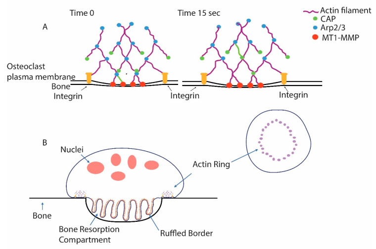
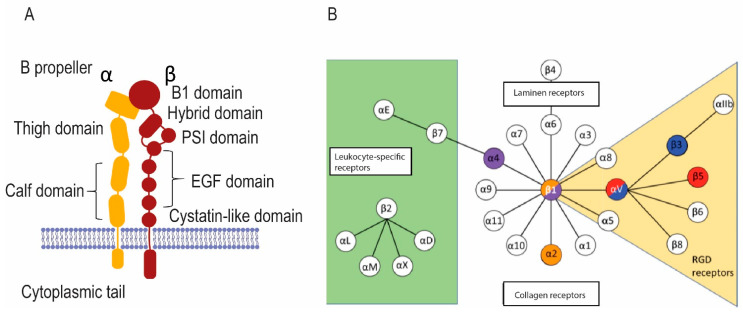
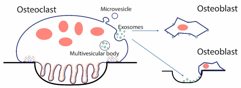
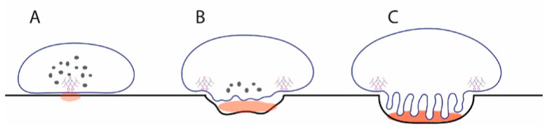
Osteoclasts differentiate from hematopoietic cells and resorb the bone in response to various signals, some of which are received directly from noncellular elements of the bone. In vitro, adherence to the bone triggers the reduction of cell-cell fusion events between osteoclasts and the activation of osteoclasts to form unusual dynamic cytoskeletal and membrane structures that are required for degrading the bone. Integrins on the surface of osteoclasts are known to receive regulatory signals from the bone matrix. Regulation of the availability of these signals is accomplished by enzymatic alterations of the bone matrix by protease activity and phosphorylation/dephosphorylation events. Other membrane receptors are present in osteoclasts and may interact with as yet unidentified signals in the bone. Bone mineral has been shown to have regulatory effects on osteoclasts, and osteoclast activity is also directly modulated by mechanical stress. As understanding of how osteoclasts and other bone cells interact with the bone has emerged, increasingly sophisticated efforts have been made to create bone biomimetics that reproduce both the structural properties of the bone and the bone's ability to regulate osteoclasts and other bone cells. A more complete understanding of the interactions between osteoclasts and the bone may lead to new strategies for the treatment of bone diseases and the production of bone biomimetics to repair defects.

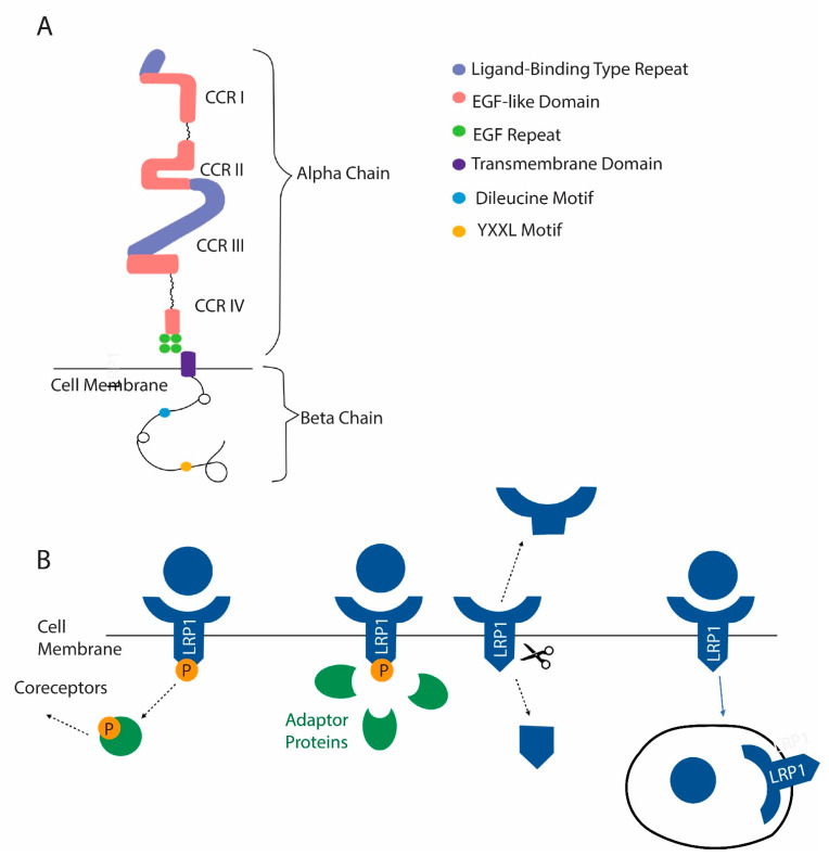
Keywords: CD44; LRP1; V-ATPase; bone remodeling; extracellular vesicles; integrins; osteopontin; synaptotagmin; vacuolar H+-ATPase.


Conflict of interest statement

The authors declare no conflict of interest.

Figures

Figure 1
Figure 1
Podosomes are the core unit of the actin ring of osteoclasts. (A) Podosomes are discrete and dynamic microfilament-based structures that are associated with the ability of cells to invade the extracellular matrix. Constant actin polymerization is triggered by the actin-related 2/3 (Arp2/3) complex, which binds pre-existing filaments and triggers new filament formation. Capping protein (CAP) quickly binds the growing actin filaments’ end and prevents further growth, limiting the size of the individual filaments. Note that one actin filament is green and that, from time 0 to 15 s, it has finished growing and been capped and been translocated in the filament network from the plasma membrane where it polymerized. Podosomes are connected with the less dense actin filament network of the cell, which gives them purchase as the filament polymerization exerts force on the membrane. Integrins bind the matrix and hold the cells tight against the podosomes pushing against the membrane. Membrane type 1-matrix metalloproteinase (MT1-MMP) associated with the membrane degrades matrix proteins. (B) In osteoclasts, many podosomes are woven into a higher-order structure, the actin ring. Force from the podosomes of the actin ring pushes the membrane into the bone, forming a tightly sealed extracellular resorption compartment. Vacuolar H+-ATPases (V-ATPases) are inserted, as vesicles fuse with the nascent ruffled plasma membrane, and then pump protons into the resorption compartment, lowering the pH, solubilizing bone mineral, and providing an environment suitable for the protease activity of cathepsin K, which is secreted into the resorption compartment and is active in acidic environments. Cathepsin K is the primary agent for the degradation of the organic matrix of the bone.
Figure 2
Figure 2
Comparison of inactive osteoclasts in glass substrate with resorbing osteoclasts in the bone. (A) Microfilaments, detected with phalloidin, of a large inactive osteoclast are integrated into podosomes that surround the periphery of the giant cell. Long arrows point to parts of the “actin belt” of podosomes. Arrowheads show small preosteoclasts in the area of the giant cell. (B) V-ATPase, detected with an anti-E-subunit antibody, shows the vesicular distribution in the giant cell, which is very flat (less than 2 microns thick). (C) The actin rings of two osteoclasts—the larger resorbing cell is outlined, and the arrowhead points to the actin ring of a smaller resorbing mononuclear osteoclast. (D) The V-ATPase is mostly packed into the ruffled borders of the resorbing cells, which are bounded by the actin rings. Scale bar equals 50 µm in (A,B) and 10 µm (C,D).
Figure 3
Figure 3
Integrins in osteoclasts bind to and detect signals in the bone matrix. (A) The general structure of integrins. Transmembrane α and β integrin proteins pair in various ways to make integrins with various specificities and signaling properties. (B) The chart shows various integrin pairs; colored integrins are found in osteoclasts.
Figure 4
Figure 4
LRP1 is a newly identified membrane receptor with a role in osteoclast function. (A) Domain structure of LRP1. (B) Mechanisms by which LRP1 functions include the activation of coreceptors after ligand binding. The activation of signaling pathways after recruitment of adapter proteins can occur in response to ligand binding. LRP1 can be cleaved, releasing a soluble extracellular domain and a cytosolic domain that can act in the nucleus. Finally, LRP1 can act as a scavenger receptor. After certain ligands are bound, they are internalized with LRP1.
Figure 5
Figure 5
EVs from osteoclasts could signal osteoblasts and other cells directly, or could bind the bone or extracellular matrix serving as matrix-associated signaling units. Osteoclasts can shed two types of EVs, exosomes, which are released when the multivesicular body fuses with the plasma membrane. Microvesicles bud directly from the plasma membrane. In either case, EVs can directly interact with target cells or can bind the bone, as shown in a resorption pit above, and other forms of extracellular matrix through membrane receptors such as integrins, and the bound form can then interact with cells.
Figure 6
Figure 6
Model for how release of calcium at initial podosome patch could stimulate vesicle fusion to generate a ruffled border. (A) Initial patch of microfilaments and V-ATPase locally lowers pH sufficiently to release soluble calcium (light brown). Most V-ATPases are in cytosolic V-ATPase storage vesicles. (B) Calcium detected by synaptotagmin VII triggers fusion of cytosolic vesicles, introducing V-ATPases into the emerging ruffled border. This pumps more protons into the resorption compartment, leading to more release of soluble calcium. (C) Over time, this process continues until the mature resorption compartment of the resorbing osteoclast is formed, leaving no V-ATPase storage vesicles in the cytosol. The osteoclasts maintain low levels of other subsets of V-ATPase in the endosomal system for housekeeping functions.

Similar articles

Cited by

References

    1. Florencio-Silva R., da Silva Sasso G.R., Sasso-Cerri E., Simões M.J., Cerri P.S. Biology of Bone Tissue: Structure, Function, and Factors That Influence Bone Cells. BioMed Res. Int. 2015;2015:421746. doi: 10.1155/2015/421746. - DOI - PMC - PubMed
    1. Clarke B. Normal Bone Anatomy and Physiology. Clin. J. Am. Soc. Nephrol. 2008;3:S131–S139. doi: 10.2215/CJN.04151206. - DOI - PMC - PubMed
    1. Einhorn T.A., Gerstenfeld L.C. Fracture healing: Mechanisms and interventions. Nat. Rev. Rheumatol. 2015;11:45–54. doi: 10.1038/nrrheum.2014.164. - DOI - PMC - PubMed
    1. McInnes I.B., Schett G. The pathogenesis of rheumatoid arthritis. N. Engl. J. Med. 2011;365:2205–2219. doi: 10.1056/NEJMra1004965. - DOI - PubMed
    1. Macedo F., Ladeira K., Pinho F., Saraiva N., Bonito N., Pinto L., Gonçalves F. Bone metastases: An overview. Oncol. Rev. 2017;11:321. doi: 10.4081/oncol.2017.321. - DOI - PMC - PubMed

MeSH terms