

Mechanical Studies of the Third Dimension in Cancer: From 2D to 3D Model
- PMID: 34576261
- PMCID: PMC8472581
- DOI: 10.3390/ijms221810098
Mechanical Studies of the Third Dimension in Cancer: From 2D to 3D Model
Abstract
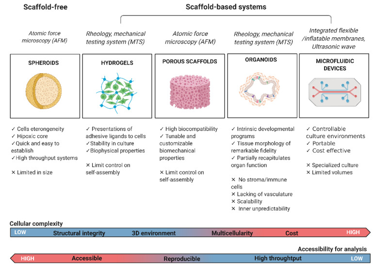
From the development of self-aggregating, scaffold-free multicellular spheroids to the inclusion of scaffold systems, 3D models have progressively increased in complexity to better mimic native tissues. The inclusion of a third dimension in cancer models allows researchers to zoom out from a significant but limited cancer cell research approach to a wider investigation of the tumor microenvironment. This model can include multiple cell types and many elements from the extracellular matrix (ECM), which provides mechanical support for the tissue, mediates cell-microenvironment interactions, and plays a key role in cancer cell invasion. Both biochemical and biophysical signals from the extracellular space strongly influence cell fate, the epigenetic landscape, and gene expression. Specifically, a detailed mechanistic understanding of tumor cell-ECM interactions, especially during cancer invasion, is lacking. In this review, we focus on the latest achievements in the study of ECM biomechanics and mechanosensing in cancer on 3D scaffold-based and scaffold-free models, focusing on each platform's level of complexity, up-to-date mechanical tests performed, limitations, and potential for further improvements.
Keywords: 3D model; biomaterials; cancer; mechanics; mechanosensing; microenvironment.
Conflict of interest statement
The authors declare no conflict of interest.
Figures
References
Publication types
MeSH terms
Substances
LinkOut - more resources
Full Text Sources
Medical
