Review
. 2021 Sep 18;22(18):10110.
doi: 10.3390/ijms221810110.

Actions and Roles of FSH in Germinative Cells

Affiliations
Review

Actions and Roles of FSH in Germinative Cells

Kaiana Recchia et al. Int J Mol Sci. .

Abstract

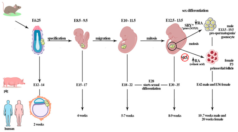
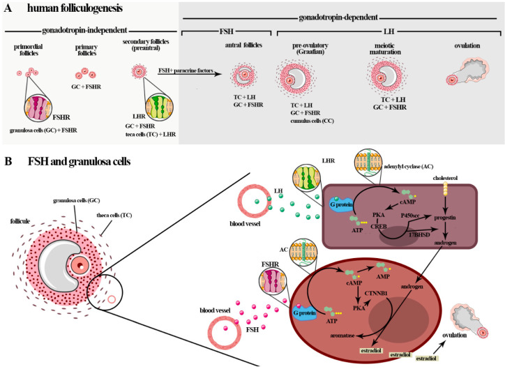
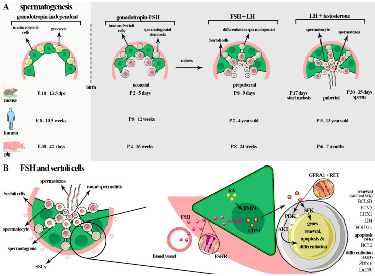
Follicle stimulating hormone (FSH) is produced by the pituitary gland in a coordinated hypothalamic-pituitary-gonadal (HPG) axis event, plays important roles in reproduction and germ cell development during different phases of reproductive development (fetal, neonatal, puberty, and adult life), and is consequently essential for fertility. FSH is a heterodimeric glycoprotein hormone of two dissociable subunits, α and β. The FSH β-subunit (FSHβ) function starts upon coupling to its specific receptor: follicle-stimulating hormone receptor (FSHR). FSHRs are localized mainly on the surface of target cells on the testis and ovary (granulosa and Sertoli cells) and have recently been found in testicular stem cells and extra-gonadal tissue. Several reproduction disorders are associated with absent or low FSH secretion, with mutation of the FSH β-subunit or the FSH receptor, and/or its signaling pathways. However, the influence of FSH on germ cells is still poorly understood; some studies have suggested that this hormone also plays a determinant role in the self-renewal of germinative cells and acts to increase undifferentiated spermatogonia proliferation. In addition, in vitro, together with other factors, it assists the process of differentiation of primordial germ cells (PGCLCs) into gametes (oocyte-like and SSCLCs). In this review, we describe relevant research on the influence of FSH on spermatogenesis and folliculogenesis, mainly in the germ cell of humans and other species. The possible roles of FSH in germ cell generation in vitro are also presented.

Keywords: germ cell line; gonadotrophin; reproduction.


Conflict of interest statement

The authors declare no conflict of interest.

Figures

Figure 1
Figure 1
The mammalian gametogenesis process starts after the primordial germ cells (PGCs) complete the migration process to the gonadal ridge. The PGCs migration window is specific for each mammal species (e.g., ~E8–9.5 days for mice, ~E15–17 days for pigs, ~E25–30 days for cows, and ~4 weeks for humans). During PGCs colonization, the gonads are undifferentiated and, morphologically, are apparently identical (bipotential period). In porcine, gonadal differentiation starts at 28 days of gestation; however, complete differentiation is observed at E30–35 days of gestation. By E42, males have gonocytes in differentiated gonads; in females, the first primordial follicle is observed at E56 d.p.c in the fetal ovary. In mice, the germ cells of embryos at day E12.5 are morphologically undifferentiated; however, initiation of sex differentiation occurs at E13.5 days; the gonocytes are found from E13.5 until birth. In humans, PGCs colonize the genital ridge at 5–7 weeks (~E37), oogonia proliferation occurs until 10 weeks when the cells enter meiotic prophase, and primordial follicles are observed at 20 weeks of gestation. Sex determination is orchestrated by the SRY gene on the Y chromosome. *The expression of the SRY gene promotes testis formation through the activation of other genes (testicular genes, e.g., other factors and genes such as SOX9) during sex differentiation. The retinoic acid (RA) plays important role when PGCs enter meiosis. The female germ cells start meiosis after RA action, differently from males, in which meiosis pathway is inhibited by the RA-degrading enzyme CYP26B1.This period is gonadotropin-independent, although Sertoli cells expresses FSHR, that starts to act after birth.
Figure 2
Figure 2
(A) Initially, the follicular growth that begins during gestation is gonadotropin-independent, although granulosa cells are influenced by FSH. The post-natal phases are gonadotrophin-dependent, and FSH influences the transition of the GCs of the larger antral follicles to the ovulatory follicles. After birth, folliculogenesis, a highly regulated process, can be classified into three phases: follicle growth, transition and maturation, and ovulation. In humans and mice, five stages of follicular development are described: primordial follicles, primary follicles, secondary follicles (preantral), antral follicles, and preovulatory follicles (Graafian). The phase of follicle transition from the preantral stage to the early antral stage and follicle growth and maturation is dependent on stimulus by FSH and LH (gonadotropin-dependent phase), which play an obligatory role in follicle differentiation, selection, and survival. (B) FSH actions in folliculogenesis, including in the induction of GCs proliferation and stimulation of GCs in the estradiol production by aromatase (CYP19A1) conversion of the androgens produced by theca cells (i.e., dehydroepiandrosterone (DHEA), androstenediol, androstenedione, and testosterone) from cholesterol in responding to LH (steroidogenesis). The interaction of the FSH-FSHR localized in the membrane surfaces of GCs activates adenylyl cyclase and stimulates the proliferation of CG cells by activating the cyclic adenosine monophosphate/protein kinase A (cAMP/PKA), mitogen-activated protein kinase/extracellular signal-regulated kinase (MAPK/ERK), and PI3K/Akt pathways. In particular, cAMP/PKA promotes the phosphorylation of cyclic AMP response element-binding (CREB) protein and other proteins that promote an increase in the expressions of genes that encode the growth factors and proteins involved in steroid hormone production and cellular growth. This gonadotropin can impede the apoptosis of GCs via the PI3K/Akt pathway.
Figure 3
Figure 3
(A) In fetal life, PGCs transform into gonocytes that remain centrally placed, surrounded by immature Sertoli cells. In mice, gonocyte development occurs before the formation at E10-13.5 d.p.c, in humans at E 8–10.5 weeks, and in pigs at E30–42 d.p.c (gonadotropin-independent). In the neonatal phase, FSH, through FSHR signaling, regulates the proliferation of the cells and the number of cells that will be had in adult life (~P2–5-day mice (postpartum), ~P8–12 weeks in humans, and ~P4–16 weeks in pigs). During the prepubertal phase, an increase in FSH occurs during the maturation of Sertoli cells and during the completion of the first cycle of sperm (~P8–10 days in mice; ~P2–4 years in humans; ~P8–24 weeks in pigs). In adult life, the spermatogenesis process starts. This is a complex process in which diploid spermatogonia self-renew, proliferate, and differentiate into haploid spermatozoa. The gonadotropins act in the early events of the spermatogenesis, before spermiogenesis, mainly in spermatogonial proliferation and meiosis. These hormones act on all phases of spermatogenesis in some species such as rodents and a specific phase of spermatogenesis in men: the maturation of type A spermatogonia to type B spermatogonia, meiosis, and spermiation. (B) In germ cells, FSH mainly influences self-renewal, proliferation, and survival of spermatogonia cells through glial cell-line-derived neurotrophic factor (GDNF) secreted by Sertoli cells. Sertoli cells secrete many factors linked to self-renewal such as GDNF and fibroblast growth factor 2 (FGF2), differentiation and proliferation of spermatogonial stem cells (SSCs), bone morphogenetic protein (BMP4), and activin A, amongst others such as KIT ligand (KL or stem cell factor—SCF), which promotes the KIT tyrosine-kinase receptor expressed by differentiated spermatogonia. GDNF induces SSC self-renew and survival through multiple pathways such as AKT/MEK, AKT, and SFK. The phosphoinositide 3-kinase (PI3K)/AKT pathway influences the self-renewing divisions of SSCs, inhibits apoptosis, and is involved in activating mTORC1 through the SFK signaling pathway. GDNF upregulates the specific SSC genes such as B cell CLL/lymphoma 6, member B (BCL6B), Ets variant gene 5 (ETV5), and Lim homeobox protein 1 (LHX1). GDNF also acts on the canonical RAS/ERK1/2 pathway, important for the proliferation and self-renewal of these cells by phosphorylation and activation of CREB1, ATF1, CREM, and c-FOS factors.

References

    1. Oduwole O.O., Peltoketo H., Huhtaniemi I.T. Role of follicle-stimulating hormone in spermatogenesis. Front. Endocrinol. 2018;9:763. doi: 10.3389/fendo.2018.00763. - DOI - PMC - PubMed
    1. Wang H.-Q., Zhang W.-D., Yuan B., Zhang J.-B. Advances in the Regulation of Mammalian Follicle-Stimulating Hormone Secretion. Animals. 2021;11:1134. doi: 10.3390/ani11041134. - DOI - PMC - PubMed
    1. Arroyo A., Kim B., Yeh J. Luteinizing Hormone Action in Human Oocyte Maturation and Quality: Signaling Pathways, Regulation, and Clinical Impact. Reprod. Sci. 2020;27:1223–1252. doi: 10.1007/s43032-019-00137-x. - DOI - PMC - PubMed
    1. Padmanabhan V., Cardoso R.C. Neuroendocrine, autocrine, and paracrine control of follicle-stimulating hormone secretion. Mol. Cell. Endocrinol. 2020;500:110632. doi: 10.1016/j.mce.2019.110632. - DOI - PMC - PubMed
    1. Patel H., Bhartiya D. Testicular Stem Cells Express Follicle-Stimulating Hormone Receptors and Are Directly Modulated by FSH. Reprod. Sci. 2016;23:1493–1508. doi: 10.1177/1933719116643593. - DOI - PubMed

MeSH terms

Substances