Review
. 2021 Sep 18;22(18):10116.
doi: 10.3390/ijms221810116.

Synthesis and Application of Silica-Coated Quantum Dots in Biomedicine

Affiliations
Review

Synthesis and Application of Silica-Coated Quantum Dots in Biomedicine

Xuan-Hung Pham et al. Int J Mol Sci. .

Abstract

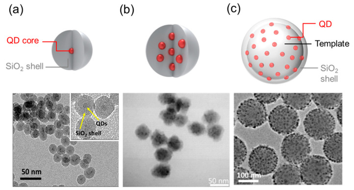
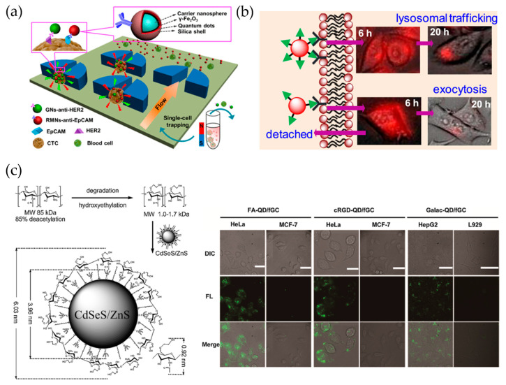
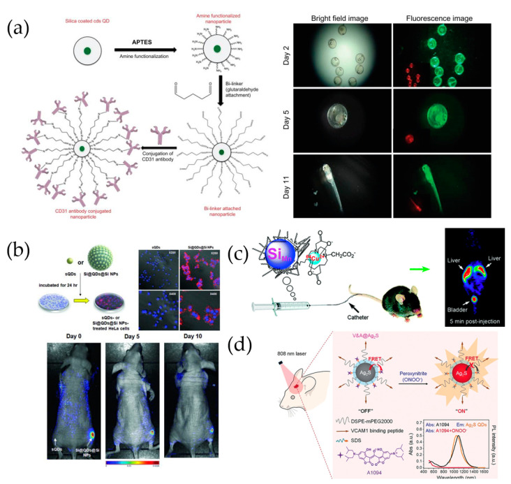
Quantum dots (QDs) are semiconductor nanoparticles with outstanding optoelectronic properties. More specifically, QDs are highly bright and exhibit wide absorption spectra, narrow light bands, and excellent photovoltaic stability, which make them useful in bioscience and medicine, particularly for sensing, optical imaging, cell separation, and diagnosis. In general, QDs are stabilized using a hydrophobic ligand during synthesis, and thus their hydrophobic surfaces must undergo hydrophilic modification if the QDs are to be used in bioapplications. Silica-coating is one of the most effective methods for overcoming the disadvantages of QDs, owing to silica's physicochemical stability, nontoxicity, and excellent bioavailability. This review highlights recent progress in the design, preparation, and application of silica-coated QDs and presents an overview of the major challenges and prospects of their application.

Keywords: bioapplication; quantum dot (QD); silica coating; silica encapsulation; surface modification.


Conflict of interest statement

The authors declare no conflict of interest.

Figures

Figure 1
Figure 1
Quantum dot (QD) classification: (a) Single QD coated SiO2 shell [13], (b) Multiply QD-doped SiO2 nanoparticle [30], (c) Template-based multi-QDs [31].
Figure 2
Figure 2
(a) Fluorescence-SERS QD-embedded silver bumpy nanoprobes, which have a SiO2@AgNS@SiO2@QD2@SiO2 structure. Forty-five different dual-modal nanoprobes were prepared from silica-coated silver bumpy nanoshells (AgNS@SiO2) with 15 different types of Raman label compounds and 3 types of QDs (red, green, and blue) [42]. (b) Mn-doped ZnS (ZnS:Mn) QDs combining dual ligands (oleylamine and octadecylamine), hydrophilic glutathione (GSH) ligands, and silica for biomedical application. Adapted with permission from ACS Appl. Nano Mater. 2020, 3, 3, 3088–3096. Copyright 2020 American Chemical Society [49]. (c) Dual-functional nanoprobe composed of an iron oxide core surrounded by QD-embedded silica NPs [44]. (d) SiO2@QDs@PDA NPs for label-free, multiplexed detection of biological molecules [47].
Figure 3
Figure 3
(a) QD-introduced self-assembly procedure into silica NPs (SiO2@QD-TACs) and cell immunolabeling [53]. (b) QD-embedded silica-coated Ag NPs (Ag@SiO2/QDs) and attached aptamer (Ag@SiO2/QDs-Apt). Tc detection using shell-isolated NP-enhanced fluorescence (SHINEF) [54]. (c) Detection of zealralenone (ZEN) and deoxynivalenol (DON) using LFIA with QD@SiO2 [55]. (d) Detection of human foreskin fibroblast exosomes using fLFA with M-QD-SNs [56].
Figure 4
Figure 4
(a) Ratiometric fluorescence detection of ascorbic acid (AA) based on the “OFF” and “ON” step of a nanoprobe that contains a blue-emitting carbon dot (CD) and red-emitting, silica-coated QDs (QDs@SiO2) [62]. (b) Fluorescence detection of serotonin (5-HT) using hybrid QDs, silica, and molecularly imprinted polymers (QDs@SiO2@MIPs) [63]. (c) Selective recognition of malachite green (MG) using MIP-coated QDs [64]. (d) Specific recognition and fluorescence quantification of bovine serum albumin (BSA) using epitope MIP (EMIP) coated QDs [65].
Figure 5
Figure 5
(a) Fabrication of IgG-QD@SiO2 conjugate via surface modification of QD@SiO2 and indirect immunoassay with IgG-QD@SiO2 conjugate [68]. Reprinted with permission from Anal. Chem. 2018, 90, 17, 10518–10526. Copyright 2018 American Chemical Society. (b) Cell uptake of QDs with different bioconjugation multivalencies [73]. Reprinted with permission from Langmuir 2019, 35, 35, 11380–11388. Copyright 2019 American Chemical Society. (c) Synthesis and use (live-cell imaging) of glycol chitosan-shelled QDs (QD/fGC) [74].
Figure 6
Figure 6
(a) Antibody conjugation onto silica-coated CdS QDs and in vivo fluorescence imaging of silica-coated CdS QD-treated embryos [75]. (b) In vivo cellular uptake of sQDs and SiO2@QDs@ SiO2 NPs [37]. (c) Use of dextran-coated silicon QDs in PET imaging and biodistribution [77]. Reprinted with permission from ACS Med. Chem. Lett. 2011, 2, 4, 285–288. Copyright 2011 American Chemical Society. (d) V&A@Ag2S probe preparation and in vivo detection of peroxynitrite [78].

References

    1. Zhang L., Zhao Z.J., Wang T., Gong J. Nano-designed semiconductors for electro- and photoelectro-catalytic conversion of carbon dioxide. Chem. Soc. Rev. 2018;47:5423–5443. doi: 10.1039/C8CS00016F. - DOI - PubMed
    1. Koo K.M., Mainwaring P.N., Tomlins S.A., Trau M. Merging new-age biomarkers and nanodiagnostics for precision prostate cancer management. Nat. Rev. Urol. 2019;16:302–317. doi: 10.1038/s41585-019-0178-2. - DOI - PubMed
    1. Lee S.H., Jun B.H. Silver nanoparticles: Synthesis and application for nanomedicine. Int. J. Mol. Sci. 2019;20:865. doi: 10.3390/ijms20040865. - DOI - PMC - PubMed
    1. Rho W.Y., Yang H.Y., Kim H.S., Son B.S., Suh J.S., Jun B.H. Recent advances in plasmonic dye-sensitized solar cells. J. Solid State Chem. 2018;258:271–282. doi: 10.1016/j.jssc.2017.10.018. - DOI
    1. Hahm E., Cha M.G., Kang E.J., Pham X.H., Lee S.H., Kim H.M., Kim D.E., Lee Y.S., Jeong D.H., Juns B.H. Multilayer Ag-embedded silica nanostructure as a surface-enhanced raman scattering-based chemical sensor with dual-function internal standards. ACS Appl. Mater. Interfaces. 2018;10:40748–40755. doi: 10.1021/acsami.8b12640. - DOI - PubMed

LinkOut - more resources