

Lipid-Based Drug Delivery Systems in Regenerative Medicine
- PMID: 34576594
- PMCID: PMC8467523
- DOI: 10.3390/ma14185371
Lipid-Based Drug Delivery Systems in Regenerative Medicine
Abstract
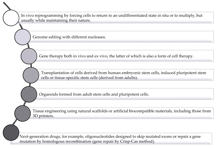
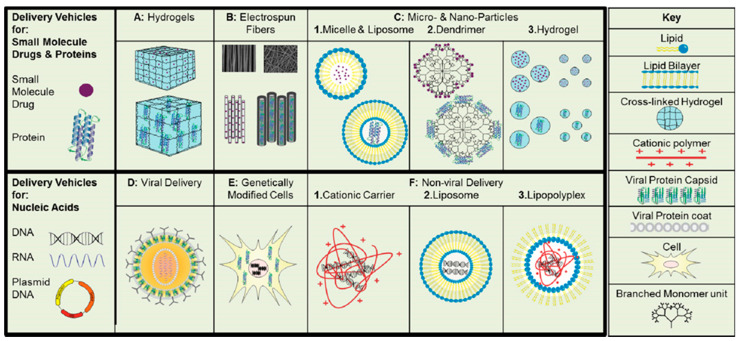
The most important goal of regenerative medicine is to repair, restore, and regenerate tissues and organs that have been damaged as a result of an injury, congenital defect or disease, as well as reversing the aging process of the body by utilizing its natural healing potential. Regenerative medicine utilizes products of cell therapy, as well as biomedical or tissue engineering, and is a huge field for development. In regenerative medicine, stem cells and growth factor are mainly used; thus, innovative drug delivery technologies are being studied for improved delivery. Drug delivery systems offer the protection of therapeutic proteins and peptides against proteolytic degradation where controlled delivery is achievable. Similarly, the delivery systems in combination with stem cells offer improvement of cell survival, differentiation, and engraftment. The present review summarizes the significance of biomaterials in tissue engineering and the importance of colloidal drug delivery systems in providing cells with a local environment that enables them to proliferate and differentiate efficiently, resulting in successful tissue regeneration.
Keywords: colloids; drug delivery systems; gene delivery; lipid nanoparticles; micelles; protein delivery; regenerative medicine.
Conflict of interest statement
The authors declare that they have no known competing financial interests or personal relationships that could have appeared to influence the work reported in this paper.
Figures
References
-
- Hasetine W. A brave new medicine. A conversation with William Haseltine. Interview by Joe Flower. Health Forum. J. 1999;42:28–65. - PubMed
-
- Passweg J.R., Baldomero H., Bader P., Bonini C., Cesaro S., Dreger P., Duarte R.F., Dufour C., Falkenburg J.H., Farge-Bancel D., et al. Hematopoietic SCT in Europe 2013: Recent trends in the use of alternative donors showing more haploidentical donors but fewer cord blood transplants. Bone Marrow. Transplant. 2015;50:476–482. doi: 10.1038/bmt.2014.312. - DOI - PMC - PubMed
Publication types
LinkOut - more resources
Full Text Sources
