. 2021 Sep 17;26(18):5648.
doi: 10.3390/molecules26185648.

Ezrin Modulates the Cell Surface Expression of Programmed Cell Death Ligand-1 in Human Cervical Adenocarcinoma Cells

Affiliations

Ezrin Modulates the Cell Surface Expression of Programmed Cell Death Ligand-1 in Human Cervical Adenocarcinoma Cells

Chihiro Tanaka et al. Molecules. .

Abstract

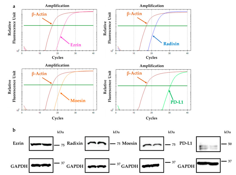
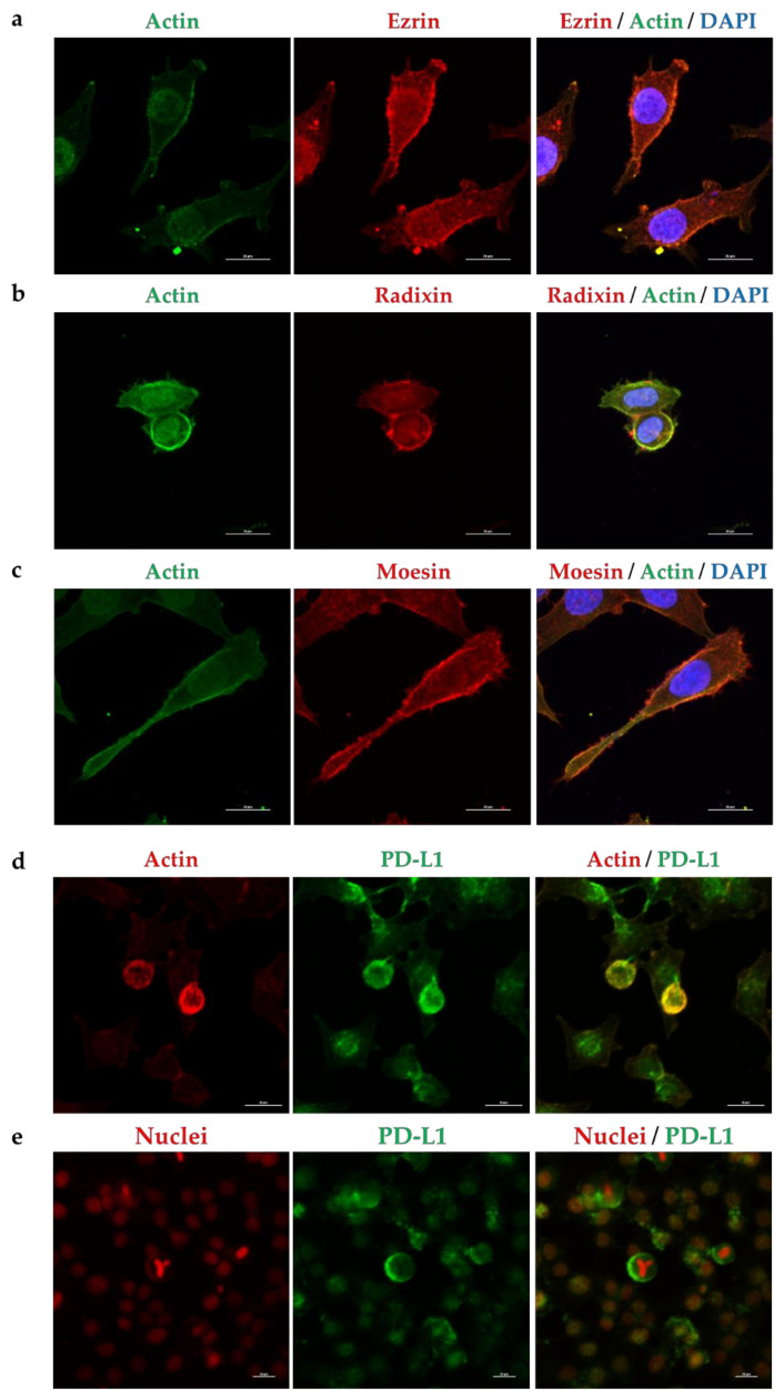
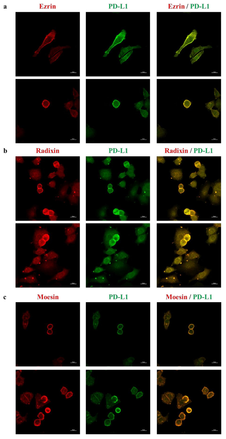
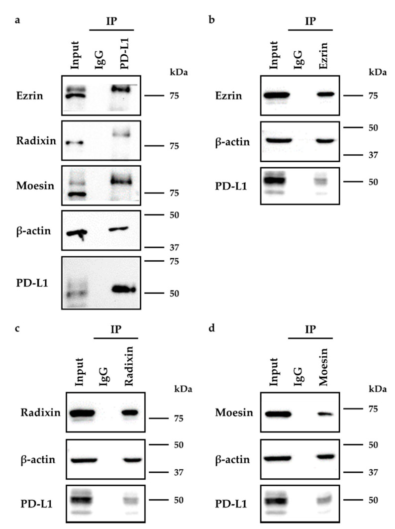
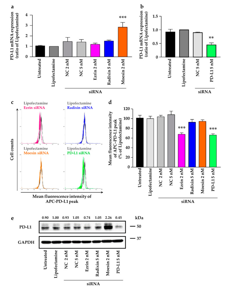
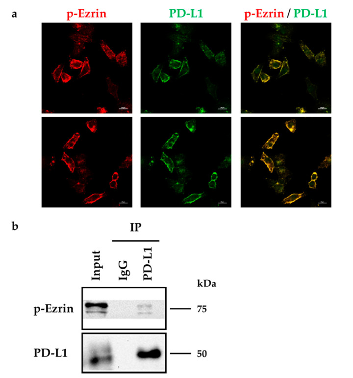
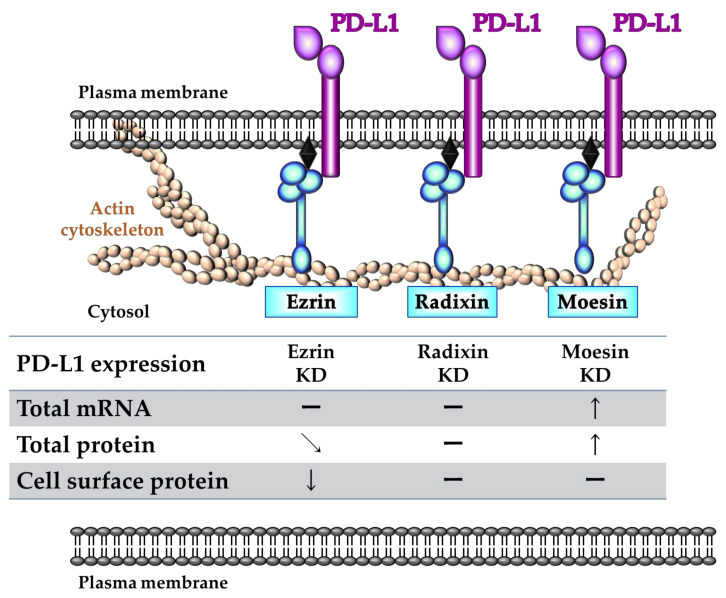
Cancer cells employ programmed cell death ligand-1 (PD-L1), an immune checkpoint protein that binds to programmed cell death-1 (PD-1) and is highly expressed in various cancers, including cervical carcinoma, to abolish T-cell-mediated immunosurveillance. Despite a key role of PD-L1 in various cancer cell types, the regulatory mechanism for PD-L1 expression is largely unknown. Understanding this mechanism could provide a novel strategy for cervical cancer therapy. Here, we investigated the influence of ezrin/radixin/moesin (ERM) family scaffold proteins, crosslinking the actin cytoskeleton and certain plasma membrane proteins, on the expression of PD-L1 in HeLa cells. Our results showed that all proteins were expressed at mRNA and protein levels and that all ERM proteins were highly colocalized with PD-L1 in the plasma membrane. Interestingly, immunoprecipitation assay results demonstrated that PD-L1 interacted with ERM as well as actin cytoskeleton proteins. Furthermore, gene silencing of ezrin, but not radixin and moesin, remarkably decreased the protein expression of PD-L1 without affecting its mRNA expression. In conclusion, ezrin may function as a scaffold protein for PD-L1; regulate PD-L1 protein expression, possibly via post-translational modification in HeLa cells; and serve as a potential therapeutic target for cervical cancer, improving the current immune checkpoint blockade therapy.

Keywords: cervical cancer; ezrin/radixin/moesin; immune check point inhibitor; programmed cell death ligand-1.


Conflict of interest statement

The authors declare no conflict of interest.

Figures

Figure 1
Figure 1
Gene and protein expression patterns of ezrin, radixin, and moesin (ERM), and programmed cell death ligand-1 (PD-L1) in HeLa cells. (a) Representative amplification curves for ezrin, radixin, moesin, and PD-L1, together with those for β-actin (internal control), in HeLa cells, as determined with reverse transcription-polymerase chain reaction (RT-PCR). (b) Western blot images of each protein in whole-cell lysates of HeLa cells. Upper panels are typical images of ezrin, radixin, moesin, and PD-L1 and lower panels are corresponding glyceraldehyde-3-phosphate dehydrogenase (GAPDH) used as internal control, all of which are shown in duplicate. Molecular weights are indicated in kDa. Data are representative of three independent experiments performed using at least three independent series of total RNA and protein extracts.
Figure 2
Figure 2
Subcellular localization of ezrin, radixin, moesin, and programmed cell death ligand-1 (PD-L1) in HeLa cells. Confocal laser scanning microscopy for subcellular localization ezrin, radixin, moesin, and PD-L1 in HeLa cells. In a three-dimensional reconstruction of optically sectioned HeLa cells, (a) ezrin, (b) radixin, and (c) moesin (red) were distributed near the plasma membrane and preferentially colocalized with actin (green), which was used as the plasma membrane maker. PD-L1 (green) was preferentially colocalized with (d) actin (red) on the plasma membrane, but not with (e) nuclei (red). Scale bars: 20 μm. All data are representative of at least three independent experiments.
Figure 3
Figure 3
Colocalization of PD-L1 with ezrin, radixin, and moesin in the plasma membrane of HeLa cells. Confocal laser scanning microscopy for subcellular localization of ezrin, radixin, moesin, and PD-L1 in HeLa cells. In a three-dimensional reconstruction of optically sectioned HeLa cells, PD-L1 (green) was highly colocalized with (a) ezrin, (b) radixin, and (c) moesin (red) on the plasma membrane. Scale bars: 20 μm. All data are representative of at least three independent experiments.
Figure 4
Figure 4
Immunoprecipitation analysis to detect the protein–protein interactions between programmed cell death ligand-1 (PD-L1) and ezrin/radixin/moesin (ERM) in HeLa cells. The whole-cell lysates of HeLa cells were immunoprecipitated with antibodies against (a) PD-L1, (b) ezrin, (c) radixin, (d) moesin, or control antibody. Western blot images of ezrin, radixin, and moesin as well as PD-L1, in addition to β-actin, in the whole-cell lysates (input) and those co-immunoprecipitated (IP) with a control IgG or each antibody, are shown. Molecular weights are indicated in kDa.
Figure 5
Figure 5
Effects of siRNAs targeting ezrin, radixin, or moesin on their total mRNA and protein expressions in HeLa cells. Cells were treated with the transfection medium (Untreated), transfection reagent (Lipofectamine), nontargeting control (NC) siRNA (2 and 5 nM), or specific siRNAs for ezrin (2 nM), radixin (5 nM), or moesin (2 nM) and then incubated for three days. (ac) Expression of each mRNA in the cells of all the treatment groups was measured via quantitative reverse transcription-polymerase chain reaction. n = 3–6, *** p < 0.001 vs. Lipofectamine. (d) Cell viability was assessed with the PrestoBlue cell viability reagent. Staurosporine was used as the positive control for inducing cell death. n = 8–16, *** p < 0.001 vs. Lipofectamine. (ad) All data are expressed as mean ± SEM and were analyzed using one-way ANOVA followed by Dunnett’s test. (e) Western blotting images of ezrin, radixin, and moesin as well as glyceraldehyde-3-phosphate dehydrogenase (GAPDH) in whole-cell lysates of HeLa cells. Molecular weights are indicated in kDa. Ratio for the chemiluminescence signal intensities of ezrin, radixin, and moesin normalized to GAPDH in each treatment group relative to Lipofectamine were shown on the respective panel.
Figure 6
Figure 6
Effects of siRNAs targeting ezrin, radixin, moesin on total mRNA, total protein, and cell surface expression of programmed cell death ligand-1 (PD-L1) in HeLa cells. Cells were treated with the transfection medium (Untreated), transfection reagent (Lipofectamine), nontargeting control (NC) siRNA (2 nM and 5 nM), or specific siRNAs for ezrin (2 nM), radixin (5 nM), moesin (2 nM), or PD-L1 (5 nM) and then incubated for 3 days. The mRNA expression of PD-L1 in cells from all treatment groups was determined via quantitative reverse transcription-polymerase chain reaction. (a) n = 3–6, *** p < 0.001 vs. Lipofectamine, (b) n = 3, ** p < 0.01 vs. Lipofectamine. All data are expressed as mean ± SEM and were analyzed using a one-way ANOVA followed by Dunnett’s test. (c) An overlay of the representative histograms for the mean fluorescence intensity of allophycocyanin (APC)-labeled PD-L1 on the surface plasma membrane of HeLa cells treated with Lipofectamine (gray line), ezrin siRNA (red line), radixin siRNA (blue line), moesin siRNA (orange line), and PD-L1 siRNA (green line), as measured by flow cytometry. (d) The calculated mean fluorescence intensities of PD-L1 relative to Lipofectamine alone on the plasma membrane surface are shown for all the treatments; n = 6, *** p < 0.001 vs. Lipofectamine. All data were expressed as the mean ± SEM and analyzed by one-way ANOVA followed by Dunnett’s test. (e) Western blotting images of PD-L1 and glyceraldehyde-3-phosphate dehydrogenase (GAPDH) in whole-cell lysates of HeLa cells. Molecular weights are indicated in kDa. Ratio for the chemiluminescence signal intensity of PD-L1 normalized to GAPDH in each treatment group relative to Lipofectamine is shown on the respective panel.
Figure 7
Figure 7
Phosphorylated ezrin is co-localized and interacted with programmed cell death ligand-1 (PD-L1) in HeLa cells. (a) Confocal laser scanning microscopy for subcellular localization of phosphorylated (p-) ezrin and PD-L1 in HeLa cells. In a three-dimensional reconstruction of optically sectioned HeLa cells, PD-L1 (green) was highly colocalized with p-ezrin (red) on the plasma membrane. Scale bars: 20 μm. Data are representative of at least three independent experiments. (b) Immunoprecipitation analysis to detect the protein–protein interactions between PD-L1 and p-ezrin in HeLa cells. The whole-cell lysates of HeLa cells were immunoprecipitated with an anti-PD-L1 antibody or its control antibody. Western blot images of p-ezrin and PD-L1 in the whole-cell lysates (input) and those co-immunoprecipitated (IP) with a control IgG or an anti-PD-L1 antibody, are shown. Molecular weights are indicated in kDa.
Figure 8
Figure 8
A proposed model illustrating the role of each ezrin/radixin/moesin (ERM) protein in the regulatory mechanism of programmed cell death ligand-1 (PD-L1) expression in HeLa cells. Ezrin regulates the plasma membrane localization of PD-L1 possibly via the protein–protein interaction, with little influences on its mRNA and total protein levels. Despite the existence of the protein–protein interaction with PD-L1, radixin and moesin might not contribute to the plasma membrane localization of PD-L1. In addition, moesin may negatively regulate PD-L1 expression at the mRNA and total protein. Therefore, among ERM proteins, ezrin plays a key role in crosslinking PD-L1 with actin cytoskeleton, resulting in a stabilization of PD-L1 in the plasma membrane of HeLa cells.

Similar articles

Cited by

References

    1. Sung H., Ferlay J., Siegel R.L., Laversanne M., Soerjomataram I., Jemal A., Bray F. Global Cancer Statistics 2020: GLOBOCAN Estimates of Incidence and Mortality Worldwide for 36 Cancers in 185 Countries. CA Cancer J. Clin. 2021;71:209–249. doi: 10.3322/caac.21660. - DOI - PubMed
    1. Liu Y., Jiang J., Liu C., Zhao W., Ma Y., Zheng Z., Zhou Q., Zhao Y. Synergistic anti-tumor effect of anti-PD-L1 antibody cationic microbubbles for delivery of the miR-34a gene combined with ultrasound on cervical carcinoma. Am. J. Transl. Res. 2021;13:988–1005. - PMC - PubMed
    1. Mauricio D., Zeybek B., Tymon-Rosario J., Harold J., Santin A.D. Immunotherapy in Cervical Cancer. Curr. Oncol. Rep. 2021;23:61. doi: 10.1007/s11912-021-01052-8. - DOI - PubMed
    1. Zhu H., Luo H., Zhang W., Shen Z., Hu X., Zhu X. Molecular mechanisms of cisplatin resistance in cervical cancer. Drug Des. Dev. Ther. 2016;10:1885–1895. doi: 10.2147/DDDT.S106412. - DOI - PMC - PubMed
    1. Shi L., Chen S., Yang L., Li Y. The role of PD-1 and PD-L1 in T-cell immune suppression in patients with hematological malignancies. J. Hematol. Oncol. 2013;6:74. doi: 10.1186/1756-8722-6-74. - DOI - PMC - PubMed

MeSH terms