

Diatom Frustule Array for Flow-Through Enhancement of Fluorescent Signal in a Microfluidic Chip
- PMID: 34577659
- PMCID: PMC8469004
- DOI: 10.3390/mi12091017
Diatom Frustule Array for Flow-Through Enhancement of Fluorescent Signal in a Microfluidic Chip
Abstract
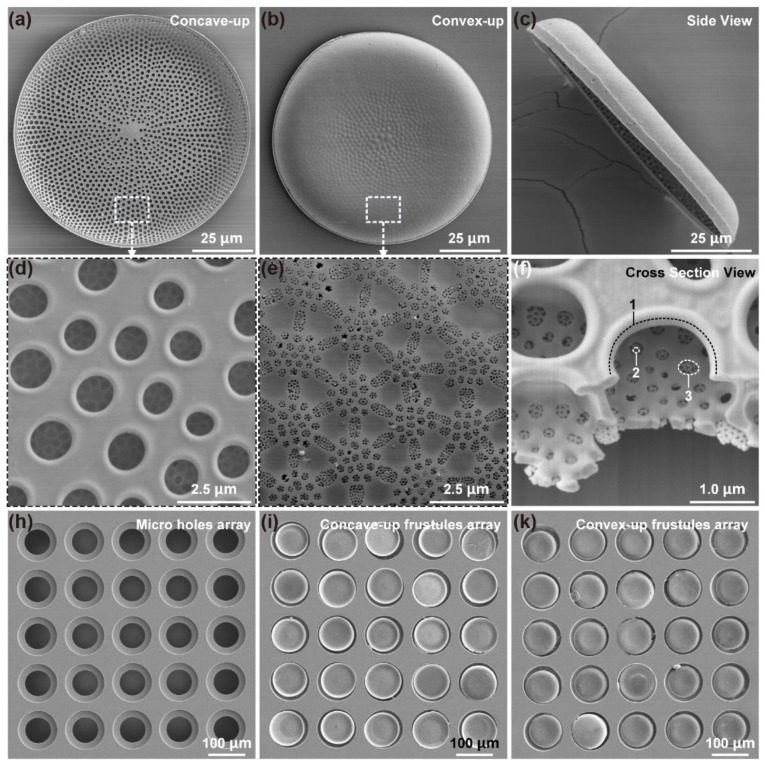
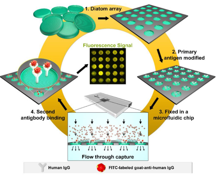
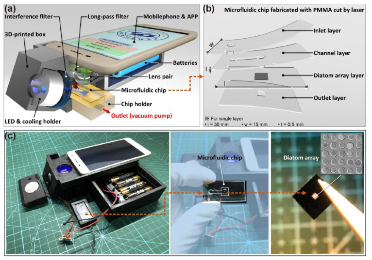
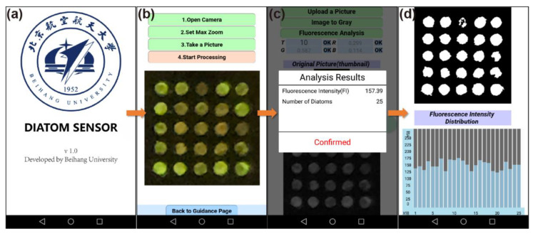
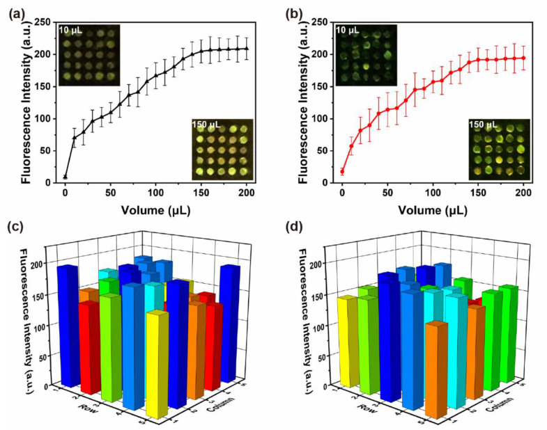
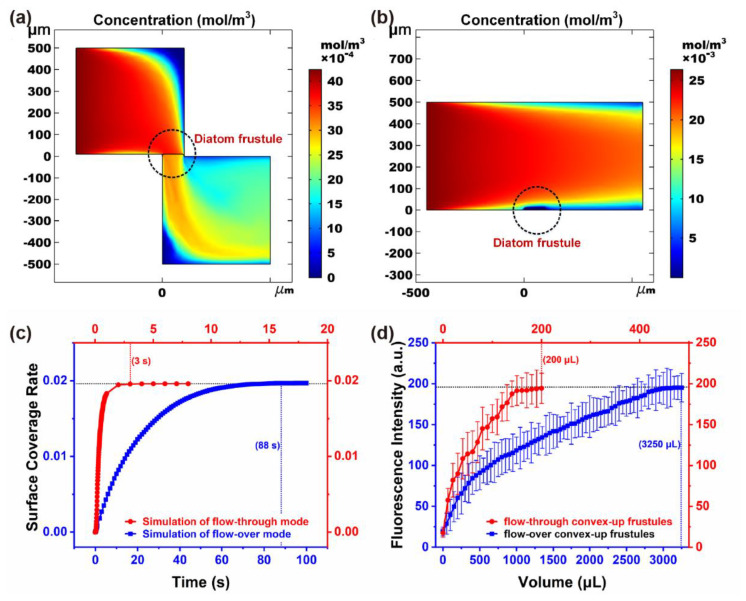
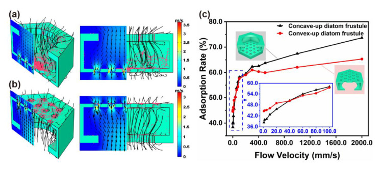
Diatom frustules are a type of natural biomaterials that feature regular shape and intricate hierarchical micro/nano structures. They have shown excellent performance in biosensing, yet few studies have been performed on flow-through detection. In this study, diatom frustules were patterned into step-through holes and bonded with silicon substrate to form an open-ended filtration array. Then they were fixed into a microfluidic chip with a smartphone-based POCT. Human IgG and FITC-labeled goat-anti-human IgG were adopted to investigate the adsorption enhancement when analyte flowed through diatom frustules. The results indicated up to 16-fold enhancement of fluorescent signal sensitivity for the flow-through mode compared with flow-over mode, at a low concentration of 10.0 μg/mL. Moreover, the maximum flow rate reached 2.0 μL/s, which resulted in a significant decrease in the testing time in POCT. The adsorption simulation results of diatom array embedded in the microchannel shows good agreement with experimental results, which further proves the filtration enrichment effect of the diatom array. The methods put forward in this study may open a new window for the application of diatom frustules in biosensing platforms.
Keywords: POCT; array; biosensing; diatom frustules; flow-through; microfluidic chip.
Conflict of interest statement
The authors declare no conflict of interest.
Figures

Similar articles
-
Study on the Hemostasis Characteristics of Biomaterial Frustules Obtained from Diatom Navicula australoshetlandica sp.Materials (Basel). 2021 Jul 5;14(13):3752. doi: 10.3390/ma14133752. Materials (Basel). 2021. PMID: 34279325 Free PMC article.
-
Gold Nanoparticle-Functionalized Diatom Biosilica as Label-Free Biosensor for Biomolecule Detection.Front Bioeng Biotechnol. 2022 May 27;10:894636. doi: 10.3389/fbioe.2022.894636. eCollection 2022. Front Bioeng Biotechnol. 2022. PMID: 35711633 Free PMC article.
-
Silica Nanowire Growth on Coscinodiscus Species Diatom Frustules via Vapor-Liquid-Solid Process.Small. 2018 Nov;14(47):e1801822. doi: 10.1002/smll.201801822. Epub 2018 Oct 4. Small. 2018. PMID: 30369025
-
Effects of abiotic factors on the nanostructure of diatom frustules-ranges and variability.Appl Microbiol Biotechnol. 2018 Jul;102(14):5889-5899. doi: 10.1007/s00253-018-9087-1. Epub 2018 May 26. Appl Microbiol Biotechnol. 2018. PMID: 29802480 Review.
-
A review on diatom biosilicification and their adaptive ability to uptake other metals into their frustules for potential application in bone repair.J Mater Chem B. 2021 Sep 14;9(34):6728-6737. doi: 10.1039/d1tb00322d. Epub 2021 Aug 3. J Mater Chem B. 2021. PMID: 34346480 Review.
Cited by
-
Limit-Defying μ-Total Analysis System: Achieving Part-Per-Quadrillion Sensitivity on a Hierarchical Optofluidic SERS Sensor.ACS Omega. 2023 May 1;8(19):17151-17158. doi: 10.1021/acsomega.3c01519. eCollection 2023 May 16. ACS Omega. 2023. PMID: 37214736 Free PMC article.
-
Diatoms: harnessing nature's microscopic marvels for biosensing and multifaceted applications.Biophys Rev. 2024 Nov 5;17(1):103-125. doi: 10.1007/s12551-024-01249-8. eCollection 2025 Feb. Biophys Rev. 2024. PMID: 40060011 Free PMC article. Review.
-
Diatom-Based Artificial Anode-Uniform Coating of Intrinsic Carbon to Enhance Lithium Storage.Materials (Basel). 2024 Sep 12;17(18):4473. doi: 10.3390/ma17184473. Materials (Basel). 2024. PMID: 39336215 Free PMC article.
References
-
- Wang Y., Zhang D., Pan J., Cai J. Key factors influencing the optical detection of biomolecules by their evaporative assembly on diatom frustules. J. Mater. Sci. 2012;47:6315–6325. doi: 10.1007/s10853-012-6554-4. - DOI
Grants and funding
LinkOut - more resources
Full Text Sources
