

Influence of the Surface Material and Illumination upon the Performance of a Microelectrode/Electrolyte Interface in Optogenetics
- PMID: 34577704
- PMCID: PMC8471589
- DOI: 10.3390/mi12091061
Influence of the Surface Material and Illumination upon the Performance of a Microelectrode/Electrolyte Interface in Optogenetics
Abstract
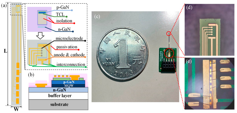
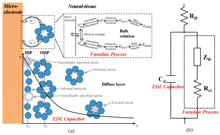
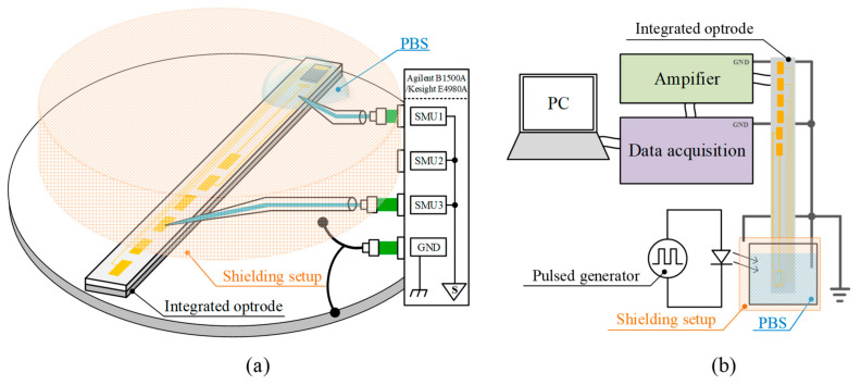
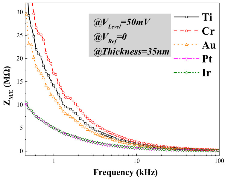
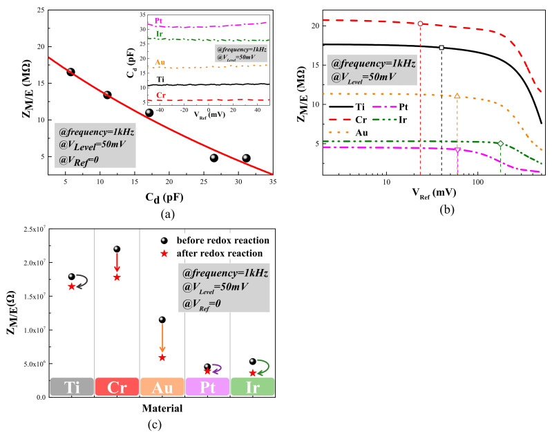
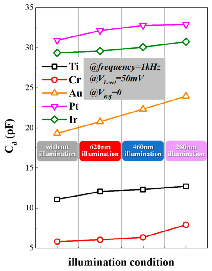
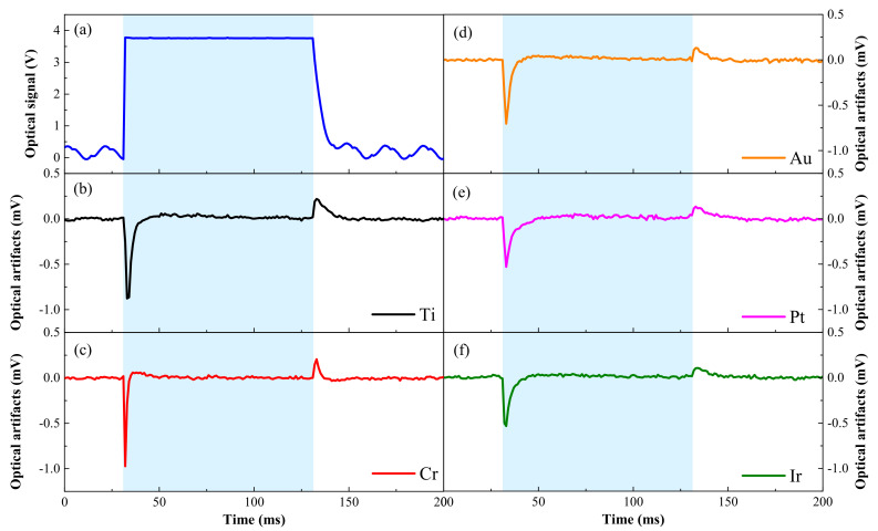
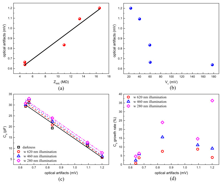
Integrated optrodes for optogenetics have been becoming a significant tool in neuroscience through the combination of offering accurate stimulation to target cells and recording biological signals simultaneously. This makes it not just be widely used in neuroscience researches, but also have a great potential to be employed in future treatments in clinical neurological diseases. To optimize the integrated optrodes, this paper aimed to investigate the influence of surface material and illumination upon the performance of the microelectrode/electrolyte interface and build a corresponding evaluation system. In this work, an integrated planar optrode with a blue LED and microelectrodes was designed and fabricated. The charge transfer mechanism on the interface was theoretically modeled and experimentally verified. An evaluation system for assessing microelectrodes was also built up. Using this system, the proposed model of various biocompatible surface materials on microelectrodes was further investigated under different illumination conditions. The influence of illumination on the microelectrode/electrolyte interface was the cause of optical artifacts, which interfere the biological signal recording. It was found that surface materials had a great effect on the charge transfer capacity, electrical stability and recoverability, photostability, and especially optical artifacts. The metal with better charge transfer capacity and electrical stability is highly possible to have a better performance on the optical artifacts, regardless of its electrical recoverability and photostability under the illumination conditions of optogenetics. Among the five metals used in our investigation, iridium served as the best surface material for the proposed integrated optrodes. Thus, optimizing the surface material for optrodes could reduce optical interference, enhance the quality of the neural signal recording for optogenetics, and thus help to advance the research in neuroscience.
Keywords: microelectrode/electrolyte interface; optical artifact; optogenetics; optrode; photostability; surface material.
Conflict of interest statement
The authors declare no conflict of interest.
Figures
Similar articles
-
Double-Sided Sapphire Optrodes with Conductive Shielding Layers to Reduce Optogenetic Stimulation Artifacts.Micromachines (Basel). 2022 Oct 27;13(11):1836. doi: 10.3390/mi13111836. Micromachines (Basel). 2022. PMID: 36363857 Free PMC article.
-
An artefact-resist optrode with internal shielding structure for low-noise neural modulation.J Neural Eng. 2020 Aug 5;17(4):046024. doi: 10.1088/1741-2552/aba41f. J Neural Eng. 2020. PMID: 32640443
-
A novel carbon tipped single micro-optrode for combined optogenetics and electrophysiology.PLoS One. 2018 Mar 7;13(3):e0193836. doi: 10.1371/journal.pone.0193836. eCollection 2018. PLoS One. 2018. PMID: 29513711 Free PMC article.
-
Recent Progress on Transparent Microelectrode-Based Soft Bioelectronic Devices for Neuroscience and Cardiac Research.ACS Appl Bio Mater. 2023 May 15;6(5):1701-1719. doi: 10.1021/acsabm.3c00131. Epub 2023 Apr 19. ACS Appl Bio Mater. 2023. PMID: 37076978 Review.
-
Multifunctional Fibers as Tools for Neuroscience and Neuroengineering.Acc Chem Res. 2018 Apr 17;51(4):829-838. doi: 10.1021/acs.accounts.7b00558. Epub 2018 Mar 21. Acc Chem Res. 2018. PMID: 29561583 Review.
Cited by
-
An Integrated Neural Optrode with Modification of Polymer-Carbon Composite Films for Suppression of the Photoelectric Artifacts.ACS Omega. 2024 Jul 17;9(30):33119-33129. doi: 10.1021/acsomega.4c04534. eCollection 2024 Jul 30. ACS Omega. 2024. PMID: 39100334 Free PMC article.
-
Double-Sided Sapphire Optrodes with Conductive Shielding Layers to Reduce Optogenetic Stimulation Artifacts.Micromachines (Basel). 2022 Oct 27;13(11):1836. doi: 10.3390/mi13111836. Micromachines (Basel). 2022. PMID: 36363857 Free PMC article.
-
Sapphire-Based Optrode for Low Noise Neural Recording and Optogenetic Manipulation.ACS Chem Neurosci. 2025 Feb 19;16(4):628-641. doi: 10.1021/acschemneuro.4c00602. Epub 2025 Feb 5. ACS Chem Neurosci. 2025. PMID: 39910858 Free PMC article.
-
Transparent neural interfaces: challenges and solutions of microengineered multimodal implants designed to measure intact neuronal populations using high-resolution electrophysiology and microscopy simultaneously.Microsyst Nanoeng. 2023 May 18;9:66. doi: 10.1038/s41378-023-00519-x. eCollection 2023. Microsyst Nanoeng. 2023. PMID: 37213820 Free PMC article. Review.
References
-
- Sahel J.-A., Boulanger-Scemama E., Pagot C., Arleo A., Galluppi F., Martel J.N., Esposti S.D., Delaux A., de Saint Aubert J.-B., de Montleau C., et al. Partial recovery of visual function in a blind patient after optogenetic therapy. Nat. Med. 2021;27:1223–1229. doi: 10.1038/s41591-021-01351-4. - DOI - PubMed
Grants and funding
- 62061160368 & 0022/2020/AFJ/This research was funded by the joint funding of the Nature Science Foundation of China (NSFC) & the Macao Science and Technology Development Fund (FDCT) of China
- 2019B010132003, 2019B010132001/Science & Technology Plan of Guangdong Province, China
- 2016YFB0400105, 2017YFB0403001/the National Key Research and Development Program
- 20167612042080001/the Zhuhai Key Technology Laboratory of Wide Bandgap Semiconductor Power Electronics, Sun Yat-sen University, China
- 088/2016/A2, 0144/2019/A3, 0022/2020/AFJ, SKL-AMSV (FDCT-funded), SKL-AMSV-ADDITIONAL FUND, SKL-AMSV(UM)-2020-2022/the Science and Technology Development Fund, Macau SAR
LinkOut - more resources
Full Text Sources
