Review
. 2021 Sep 10;57(9):951.
doi: 10.3390/medicina57090951.

The Clamshell Osteotomy for Diaphyseal Malunion in Deformity Correction and Fracture Surgery

Affiliations
Review

The Clamshell Osteotomy for Diaphyseal Malunion in Deformity Correction and Fracture Surgery

Kevin F Purcell et al. Medicina (Kaunas). .

Abstract

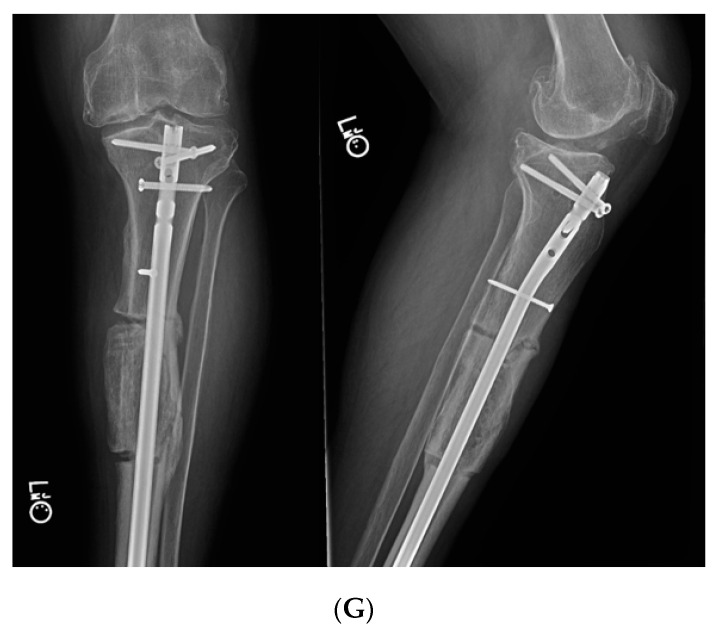
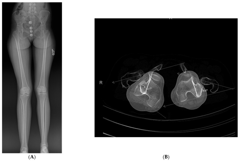
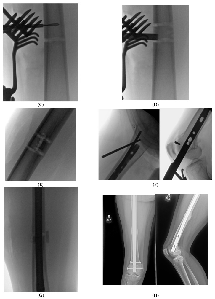
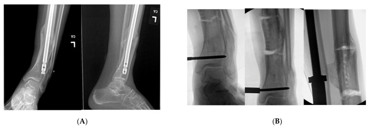
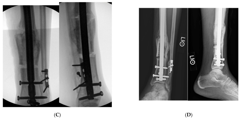
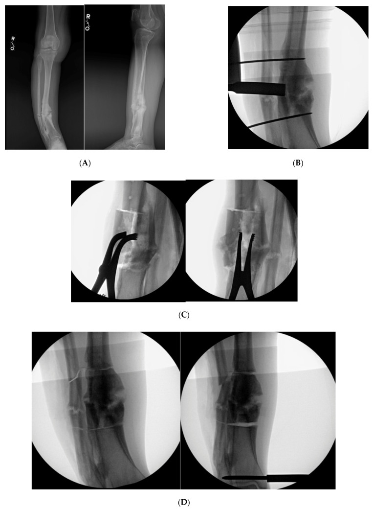
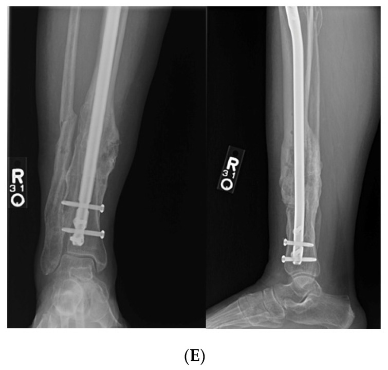
Diaphyseal malunion poses a great challenge for the orthopedic surgeon, and an inundation of morbidity for the patient. Diaphyseal malunion can cause altered gait, adjacent joint osteoarthritis and body dissatisfaction. This problem is fraught with complications without surgical intervention. There is a myriad of options for the management of a diaphyseal malunion. The clamshell osteotomy was engendered to ameliorate the difficulty in managing this issue. This technique is a viable option to correct diaphyseal malunion about the femur and tibia. Recently, the indications of a clamshell osteotomy have been expanded to function as a derotational or shortening osteotomy.

Keywords: clamshell; deformity; diaphyseal; femur; fracture; malunion; nonunion; osteotomy; tibia.


Conflict of interest statement

All authors declare no conflict of interest with regards to this publication.

Figures

Figure 1
Figure 1
Illustration of femur with bicortical drill holes in the mid axial line of the malunited segment and the perpendicular cut at the proximal and distal aspect of the malunion. (Reprinted with permission from Russell et al. (2009). Copyright 2009 The Journal of Bone and Joint Surgery (JBJS), Inc.).
Figure 2
Figure 2
(A): An osteotome is used to complete the longitudinal cut after the bicortical drill holes. (Reprinted with permission from Russell et al. (2009). Copyright 2009 The Journal of Bone and Joint Surgery (JBJS), Inc.). (B): The saw is used to create the transverse bone cuts at the proximal and distal aspect of the malunited segment. (Reprinted with permission from Russell et al. (2009). Copyright 2009 The Journal of Bone and Joint Surgery (JBJS), Inc.). (C): The osteotome is used after to open the osteotomy similar to a clamshell. A lamina spreader can be utilized as well to open the osteotomy. (Reprinted with permission from Russell et al. (2009). Copyright 2009 The Journal of Bone and Joint Surgery (JBJS), Inc.).
Figure 3
Figure 3
Illustration demonstration the orientation of the tibial clamshell osteotomy. Notice the osteotomy is parallel to the medial face of the tibia. The surgeon must ensure that he/she is not creating a unicortical osteotomy when creating the longitudinal cut of the tibia. (Reprinted with permission from Russell et al. (2009). Copyright 2009 The Journal of Bone and Joint Surgery (JBJS), Inc.).
Figure 4
Figure 4
(A): Full length standing radiographs illustrating a left tibia diaphyseal malunion. (B): AP and lateral of tibia showing a diaphyseal malunion. (C): Intraoperative fluoro demonstrating bicortical drill holes parallel to medial face of the tibia. This is prior to the utilization of the osteotome and the perpendicular saw cut. Remember to remove retractors prior to the reaming process. (D): Intraoperative fluoro illustrating the clamshell osteotomy. Notice secondary fracture line propagated during the osteotomy (red arrow). (E): The reamer was pushed pass the osteotomy zone during the reaming process. This is to prevent iatrogenic injury to neurovascular structures. (F): AP and lateral fluoroscopic images demonstrating improved alignment after clamshell osteotomy and implantation of intramedullary nail. Poller screws may be required to assist with reduction. However, the poller screw in this scenario was inserted for definitive fixation to prevent any endosteal motion of intramedullary nail. (G): AP and lateral XR demonstrating osseous union at 3 month follow up.
Figure 4
Figure 4
(A): Full length standing radiographs illustrating a left tibia diaphyseal malunion. (B): AP and lateral of tibia showing a diaphyseal malunion. (C): Intraoperative fluoro demonstrating bicortical drill holes parallel to medial face of the tibia. This is prior to the utilization of the osteotome and the perpendicular saw cut. Remember to remove retractors prior to the reaming process. (D): Intraoperative fluoro illustrating the clamshell osteotomy. Notice secondary fracture line propagated during the osteotomy (red arrow). (E): The reamer was pushed pass the osteotomy zone during the reaming process. This is to prevent iatrogenic injury to neurovascular structures. (F): AP and lateral fluoroscopic images demonstrating improved alignment after clamshell osteotomy and implantation of intramedullary nail. Poller screws may be required to assist with reduction. However, the poller screw in this scenario was inserted for definitive fixation to prevent any endosteal motion of intramedullary nail. (G): AP and lateral XR demonstrating osseous union at 3 month follow up.
Figure 5
Figure 5
(A): Full length standing radiograph. There is not a diaphyseal malunion; the patient has a 2 cm LLD, and rotational malalignment. The clamshell osteotomy was used to shorten and correct the rotational malalignment. (B): Computed tomography anteversion study demonstrating a 52° degree femoral anteversion and 30° femoral anteversion of the right and left lower extremity, respectively. (C): Intraoperative fluoro view showing bicortical drill holes being created in a diaphyseal segment measuring 2 cm. (D): An osteotome is used after the bicortical drill holes. Subsequently, a saw was used for the perpendicular cuts at the proximal and distal aspect of the osteotomy to create the clamshell. (E): Lateral fluoro view illustrating the clamshell osteotomy. No secondary fracture lines were propagated during this osteotomy. (F): 2.0 kirschner wire was placed in the proximal and distal aspect to be used as reference points when correcting the rotational malalignment. (G): AP fluoroscopic view after the clamshell segment was mobilized, and rotational malalignment was corrected. Her right knee was taken through range of motion after surgery to ensure there was not any patellofemoral maltracking. (H): AP and lateral femur XR demonstrating osseous healing of osteotomy at 6 months follow up.
Figure 5
Figure 5
(A): Full length standing radiograph. There is not a diaphyseal malunion; the patient has a 2 cm LLD, and rotational malalignment. The clamshell osteotomy was used to shorten and correct the rotational malalignment. (B): Computed tomography anteversion study demonstrating a 52° degree femoral anteversion and 30° femoral anteversion of the right and left lower extremity, respectively. (C): Intraoperative fluoro view showing bicortical drill holes being created in a diaphyseal segment measuring 2 cm. (D): An osteotome is used after the bicortical drill holes. Subsequently, a saw was used for the perpendicular cuts at the proximal and distal aspect of the osteotomy to create the clamshell. (E): Lateral fluoro view illustrating the clamshell osteotomy. No secondary fracture lines were propagated during this osteotomy. (F): 2.0 kirschner wire was placed in the proximal and distal aspect to be used as reference points when correcting the rotational malalignment. (G): AP fluoroscopic view after the clamshell segment was mobilized, and rotational malalignment was corrected. Her right knee was taken through range of motion after surgery to ensure there was not any patellofemoral maltracking. (H): AP and lateral femur XR demonstrating osseous healing of osteotomy at 6 months follow up.
Figure 6
Figure 6
(A): AP and lateral tibial XR demonstrating failure of tibial nail with valgus malunion. Notice there are not any distal interlocking screws. (B): Intraoperative fluoro views demonstrating medial universal distractor being used to assist with deformity correction, and maintain alignment during intramedullary nailing. (C): Intraoperative views demonstrating tibial nail and fibular plate after clamshell and fibular osteotomies. (D): AP and lateral 3-month post operative follow up XRs demonstrating healed clamshell osteotomy.
Figure 6
Figure 6
(A): AP and lateral tibial XR demonstrating failure of tibial nail with valgus malunion. Notice there are not any distal interlocking screws. (B): Intraoperative fluoro views demonstrating medial universal distractor being used to assist with deformity correction, and maintain alignment during intramedullary nailing. (C): Intraoperative views demonstrating tibial nail and fibular plate after clamshell and fibular osteotomies. (D): AP and lateral 3-month post operative follow up XRs demonstrating healed clamshell osteotomy.
Figure 7
Figure 7
(A): AP and lateral XR demonstration varus non-union deformity with segmental fibular fracture. (B): Intraoperative fluro view with threaded k-wire at proximal and distal aspect of malunited segment. A core reamer is being used to create the sequential bicortical drill holes. A core reamer can be used if the malunited segment is significantly larger than a 3.5 drill bit. (C): Note the lamina spreaders being utilized to open the osteotomized clamshell segment. (D): Medial universal distractor being utilized to assist with deformity correction. The distractor can be left in place during the nailing procedure. (E): AP and lateral 3-month postoperative radiographs demonstrating healed clamshell and fibular osteotomies.
Figure 7
Figure 7
(A): AP and lateral XR demonstration varus non-union deformity with segmental fibular fracture. (B): Intraoperative fluro view with threaded k-wire at proximal and distal aspect of malunited segment. A core reamer is being used to create the sequential bicortical drill holes. A core reamer can be used if the malunited segment is significantly larger than a 3.5 drill bit. (C): Note the lamina spreaders being utilized to open the osteotomized clamshell segment. (D): Medial universal distractor being utilized to assist with deformity correction. The distractor can be left in place during the nailing procedure. (E): AP and lateral 3-month postoperative radiographs demonstrating healed clamshell and fibular osteotomies.

References

    1. Santoro D., Tantavisut S., Aloj D., Karam M.D. Diaphyseal osteotomy after post-traumatic malalignment. Curr. Rev. Musculoskelet. Med. 2014;7:312–322. doi: 10.1007/s12178-014-9244-9. - DOI - PMC - PubMed
    1. Pires R.E., Gausden E.B., Sanchez G.T., Wajnsztejn A., Teixeira T.M., da Silva Soares M., Pesantez R., Lorich D.G. Clamshell Osteotomy for Acute Fractures in the Malunion Setting: A Technical Note. J. Orthop. Trauma. 2018;32:e415–e420. doi: 10.1097/BOT.0000000000001257. - DOI - PubMed
    1. Russell G.V., Graves M.L., Archdeacon M.T., Barei D.P., Brien G.A., Jr., Porter S.E. The clamshell osteotomy: A new technique to correct complex diaphyseal malunions. J. Bone Jt. Surg. Am. 2009;91:314–324. doi: 10.2106/JBJS.H.00158. - DOI - PubMed
    1. Russell G.V., Graves M.L., Archdeacon M.T., Barei D.P., Brien G.A., Jr., Porter S.E. The clamshell osteotomy: A new technique to correct complex diaphyseal malunions: Surgical technique. J. Bone Jt. Surg. Am. 2010;92((Suppl. S1)):158–175. doi: 10.2106/JBJS.I.01328. - DOI - PubMed
    1. Giannoudis P.V., Christoph Pape H., Schütz M. Antegrade Nailing Approach with Piriformis Entry Point. [(accessed on 29 June 2021)]. Available online: https://surgeryreference.aofoundation.org/orthopedic-trauma/adult-trauma....