

Complications of Intra-Arterial tPA for Iatrogenic Branch Retinal Artery Occlusion: A Case Report through Multimodal Imaging and Literature Review
- PMID: 34577886
- PMCID: PMC8464858
- DOI: 10.3390/medicina57090963
Complications of Intra-Arterial tPA for Iatrogenic Branch Retinal Artery Occlusion: A Case Report through Multimodal Imaging and Literature Review
Abstract
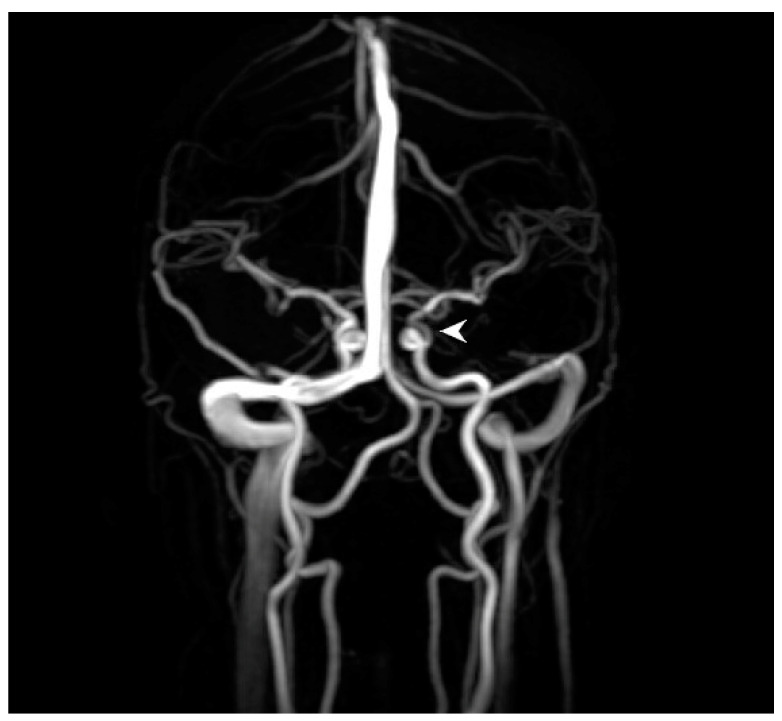
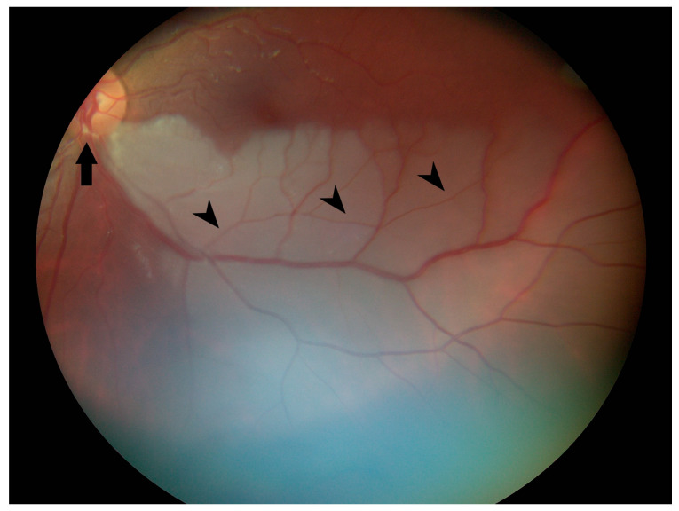
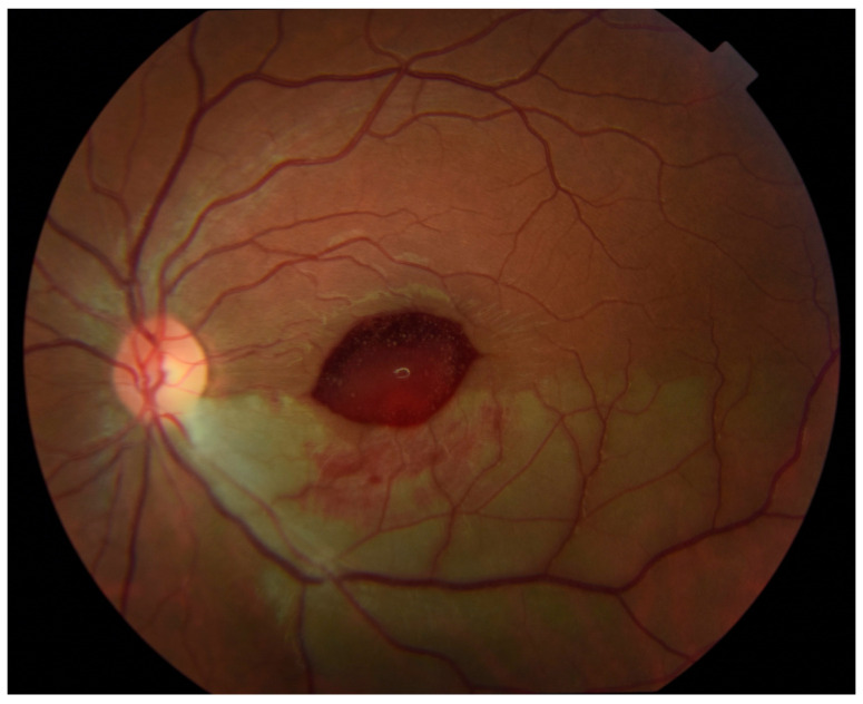
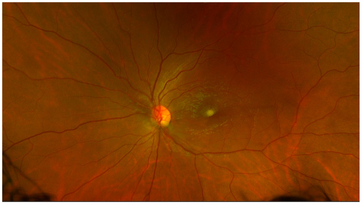
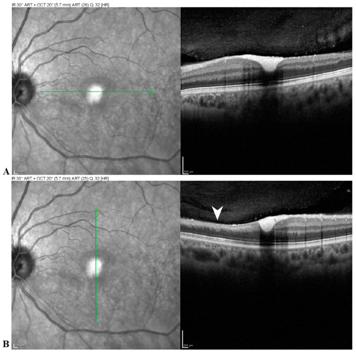
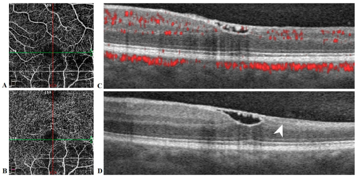
Background and Objectives: To document, through multimodal imaging, the post-procedural clinical course and visual outcome of a patient who received intra-arterial tissue plasminogen activator (tPA) for acute iatrogenic branch retinal artery occlusion (BRAO), and to review the literature and guidelines regarding the use of tPA for retinal arterial occlusions. Methods: A 28-year-old female patient who sustained an iatrogenic BRAO and subsequently received intra-arterial tPA was followed through her post-interventional course of 3 months with serial exams and multimodal imaging, including color fundus photography, visual field testing, spectral domain optical coherence tomography (SD-OCT), and OCT angiography (OCT-A). Results: A patient with history of left internal cerebral artery (ICA) aneurysm and baseline visual acuity (VA) of 20/20 developed an acutely symptomatic BRAO after undergoing a neuroendovascular procedure and was acutely treated with tPA through the left ophthalmic artery. At two weeks follow-up, a central posterior pole hemorrhage was noted although VA was preserved. A superior altitudinal defect was shown on automated perimetry. VA dropped to 20/50 at 7 weeks follow-up and hyperreflective material deep to the attachment between the posterior hyaloid and the internal limiting membrane (ILM) consistent with hemorrhage was noted on SD-OCT. At 11 weeks follow-up, VA returned to 20/20, SD-OCT revealed a membrane bridging the foveal depression, OCT-A showed decreased vascularity in the inferior macula, and the visual field defect was stable by automated perimetry. Conclusions: Intraocular hemorrhage is a possible complication of intra-arterial tPA administration for BRAO, and a careful analysis of risks, benefits, and goals of this procedure must be considered by both provider and patient before such intervention.
Keywords: optical coherence tomography angiography; retinal artery occlusion; spectral domain optical coherence tomography; tPA.
Conflict of interest statement
The authors declare no conflict of interest.
Figures
Similar articles
-
PARACENTRAL ACUTE MIDDLE MACULOPATHY AS A PRESENTING SIGN OF CENTRAL RETINAL ARTERY OCCLUSION IN SICKLE CELL DISEASE TREATED WITH TISSUE PLASMINOGEN ACTIVATOR.Retin Cases Brief Rep. 2022 Sep 1;16(5):553-557. doi: 10.1097/ICB.0000000000001033. Retin Cases Brief Rep. 2022. PMID: 32618900
-
[Spectral domain OCT in eyes with retinal artery occlusion].J Fr Ophtalmol. 2012 Oct;35(8):606-13. doi: 10.1016/j.jfo.2012.04.008. Epub 2012 Jul 21. J Fr Ophtalmol. 2012. PMID: 22819341 French.
-
Intra-arterial thrombolysis for retinal artery occlusion: the Calgary experience.Can J Neurol Sci. 2005 Nov;32(4):507-11. Can J Neurol Sci. 2005. PMID: 16408583
-
Sequential bilateral retinal artery occlusions with promising visual prognosis in a diabetic patient: a case report and literature review.BMC Ophthalmol. 2025 Jun 2;25(1):331. doi: 10.1186/s12886-025-04166-w. BMC Ophthalmol. 2025. PMID: 40457210 Free PMC article. Review.
-
Transient branch retinal artery occlusion in a 15-year-old girl and review of the literature.Biomed Pap Med Fac Univ Palacky Olomouc Czech Repub. 2015 Sep;159(3):508-11. doi: 10.5507/bp.2015.031. Epub 2015 Jul 3. Biomed Pap Med Fac Univ Palacky Olomouc Czech Repub. 2015. PMID: 26160228 Review.
Cited by
-
Vascular Repair by Grafting Based on Magnetic Nanoparticles.Pharmaceutics. 2022 Jul 8;14(7):1433. doi: 10.3390/pharmaceutics14071433. Pharmaceutics. 2022. PMID: 35890328 Free PMC article. Review.
-
Fovea Sparing Branch Retinal Artery Occlusions Imaged with Optical Coherence Tomography Angiography: Two Case Reports.Case Rep Ophthalmol. 2025 Mar 12;16(1):274-280. doi: 10.1159/000543742. eCollection 2025 Jan-Dec. Case Rep Ophthalmol. 2025. PMID: 40248824 Free PMC article.
References
-
- Sacco R.L., Kasner S.E., Broderick J.P., Caplan L.R., Connors J.J., Culebras A., Elkind M.S.V., George M.G., Hamdan A.D., Higashida R.T., et al. An Updated Definition of Stroke for the 21st Century. A Statement for Healthcare Professionals From the American Heart Association/American Stroke Association. Stroke. 2013;44:2064–2089. doi: 10.1161/STR.0b013e318296aeca. - DOI - PMC - PubMed
Publication types
MeSH terms
Substances
Grants and funding
LinkOut - more resources
Full Text Sources
Miscellaneous
