. 2021 Sep 18;57(9):985.
doi: 10.3390/medicina57090985.

Launay's External Carotid Vein

Affiliations

Launay's External Carotid Vein

Mihaela Daniela Manta et al. Medicina (Kaunas). .

Abstract

Background and Objectives: Launay's external carotid vein (ECV) is poorly represented in the anatomical literature, although it is an occasional satellite of the external carotid artery (ECA). We aimed to establish the incidence and morphology of the ECV. Materials and Methods: One hundred computed tomography angiograms were investigated, and ECVs were documented anatomically, when found. Results: Launay's vein was found in 3/200 sides (1.5%) in a male and two female cases. In two of these cases, the ECV was a replaced variant of the anterior division of the retromandibular vein (RMV), and the facial vein (FV) ended in the external jugular vein. In the third case with the ECV, the RMV was absent and the common FV that resulted from that ECV and the FV drained into the internal jugular vein. The ECV could also appear as an accessory RMV, not just as a replaced one. Additional variants were found, such as fenestration of the external jugular vein (EJV), the extracondylar vein draining the deep temporal veins and an arterial occipitoauricular trunk. Conclusions: Surgical dissections of the ECA in the retromandibular space should carefully observe an ECV to avoid unwanted haemorrhagic events. Approaches of the neck of the mandible should also carefully distinguish the consistent extracondylar veins.

Keywords: carotid artery; deep temporal veins; extracondylar vein; jugular vein; mandible; occipitoauricular trunk; parotid; retromandibular.


Conflict of interest statement

The authors declare no conflict of interest.

Figures

Figure 1
Figure 1
Right postero-infero-lateral view of the retromandibular fossa. Three-dimensional volume rendering and drawing (the branches of the external carotid artery in the carotid triangle were omitted). 1. Common carotid artery; 2. internal carotid artery; 3. internal jugular vein; 4. fenestrated external jugular vein; 5. thyro-linguo-facial trunk; 6. external carotid vein (Launay’s vein); 7. antero-medial branch of the retromandibular vein; 8. external carotid artery; 9. posterior auricular artery; 10.occipital artery; 11. posterior auricular vein; 12. posterior pharyngeal trunk; 13. superficial temporal artery; 14. transverse facial artery; 15. deep limb of the fenestrated retromandibular vein; 16. superficial limb of the fenestrated retromandibular vein; 17. postero-lateral branch of the retromandibular vein. Also included are the facial vein (18), which drains in the anterior arm of the fenestrated external jugular vein, and a submental tributary (19) of the facial vein.
Figure 2
Figure 2
Right infero-lateral view of the retromandibular fossa and carotid triangle. Three-dimensional volume rendering and drawing. 1. Common carotid artery; 2. internal jugular vein; 3. internal carotid artery; 4. external jugular vein; 5. terminal end of the facial vein; 6. occipital artery; 7. styloid process; 8. superficial temporal artery and vein; 9. maxillary veins; 10. retromandibular vein; 11. Launay’s external carotid vein; 12. facial artery; 13. external carotid artery; 14. thyro-lingual venous trunk; 15. lingual artery; 16. superior thyroid artery.
Figure 3
Figure 3
Right postero-infero-lateral view of the retromandibular fossa. Three-dimensional volume rendering and drawing. 1. Internal jugular vein; 2. external carotid artery; 3. Launay’s external carotid vein; 4. occipitoauricular trunk; 5. retrocondylar vein; 6. occipital artery; 7. posterior auricular artery; 8. internal carotid artery, styloid process; 9. superficial temporal artery; 10. superficial temporal vein; 11. deep temporal venous trunk; 12. communicating vein with the maxillary vein; 13. extracondylar vein; 14.maxillary vein; 15. intraparotid communicating vein; 16. external jugular vein (interrupted); 17. facial artery; 18. lingual artery; 19. hyoid bone; 20. proper facial vein; 21. common facial vein.
Figure 4
Figure 4
Details of the anatomic variations in Case #3. (A). Three-dimensional volume rendering, medial view of the right mandibular ramus. (B). Axial reconstructed slice through the right mandibular notch. (C). Three-dimensional volume rendering, postero-infero-lateral view of the right mandibular ramus. (D). Three-dimensional volume rendering, anterior view of Launay’s vein. 1. Maxillary vein; 2. maxillary artery; 3. extracondylar vein; 4. deep temporal venous trunk; 5. deep temporal veins; 6. communicating vein between the extracondylar and maxillary veins; 7. buccal vein; 8. temporal muscle; 9. lateral pterygoid muscle; 10. superficial temporal vein; 11. superficial temporal artery; 12. retrocondylar vein; 13. Launay’s external carotid vein; 14. occipitoauriaular trunk; 15. external carotid artery; 16. internal jugular vein; 17. anastomosis of the external carotid and external jugular veins; 18. external jugular vein; 19. proper facial vein; 20. facial artery; 21. common facial vein; 22. neck of mandible; 23. styloid process; 24. internal carotid artery.
Figure 5
Figure 5
Schematic representation of successive embryonic stages of venous development. (A). 10 mm embryo; (B).18 mm embryo; (C). 40 mm embryo. 1. Precardinal vein; 2. primitive maxillary vein; 3. ventral pharyngeal vein; 4. linguofacial vein; 5. retromandibular vein; 6. proper facial vein; 7. common facial vein; 8. internal jugular vein; 9. external jugular vein; 10. posterior division of the retromandibular vein; 11. posterior auricular vein; 12. anterior division of the retromandibular vein; 13. posterior connection of the external jugular vein; 14. anterior connection of the external jugular vein.
Figure 6
Figure 6
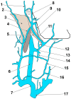
Drawing, modified after Rouvière and Delmas (1985), depicting the veins on the right side of the neck. 1. Posterior auricular vein; 2. superficial occipital vein; 3. sternocleidomastoid m.; 4. external jugular vein; 5. posterior belly of digastric m.; 6. internal jugular vein; 7. right brachiocephalic vein; 8. superficial temporal vein; 9. maxillary vein; 10. external carotid vein of Launay; 11. intraparotid communicating vein; 12. lingual vein; 13. thyro-linguo-facial trunk; 14. superior thyroid vein; 15. anterior jugular vein; 16. jugular venous arch; 17. superior vena cava.

References

    1. Laing M.R., McKerrow W.S. Intraparotid anatomy of the facial nerve and retromandibular vein. BJS. 2005;75:310–312. doi: 10.1002/bjs.1800750405. - DOI - PubMed
    1. Dalip D., Iwanaga J., Loukas M., Oskouian R.J., Tubbs R.S. Review of the Variations of the Superficial Veins of the Neck. Cureus. 2018;10:e2826. doi: 10.7759/cureus.2826. - DOI - PMC - PubMed
    1. Poletti A.M., Imparato S., Signorelli G.C., Cugini G., Colombo G. The multiplanar analysis of the retromandibular vein in surgical planning for parotid gland tumors. Eur. Arch. Oto-Rhino-Laryngol. 2018;275:1587–1593. doi: 10.1007/s00405-018-4953-0. - DOI - PubMed
    1. Imaizumi A., Kuribayashi A., Okochi K., Ishii J., Sumi Y., Yoshino N., Kurabayashi T. Differentiation between superficial and deep lobe parotid tumors by magnetic resonance imaging: Usefulness of the parotid duct criterion. Acta Radiol. 2009;50:806–811. doi: 10.1080/02841850903049358. - DOI - PubMed
    1. Henle J. Handbuch der Systematischen Anatomie des Menschen von J. Henle: Handbuch der Gefässlehre des Menschen. Friedrich Vieweg und Sohn; Braunschweig, Germany: 1868.

LinkOut - more resources