

Polyurethane-Nanolignin Composite Foam Coated with Propolis as a Platform for Wound Dressing: Synthesis and Characterization
- PMID: 34578092
- PMCID: PMC8473208
- DOI: 10.3390/polym13183191
Polyurethane-Nanolignin Composite Foam Coated with Propolis as a Platform for Wound Dressing: Synthesis and Characterization
Abstract
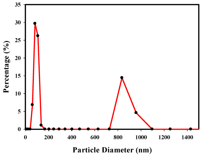
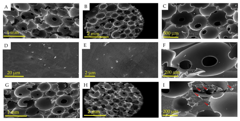
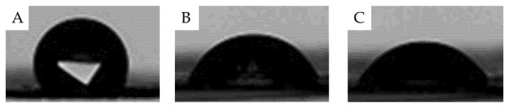
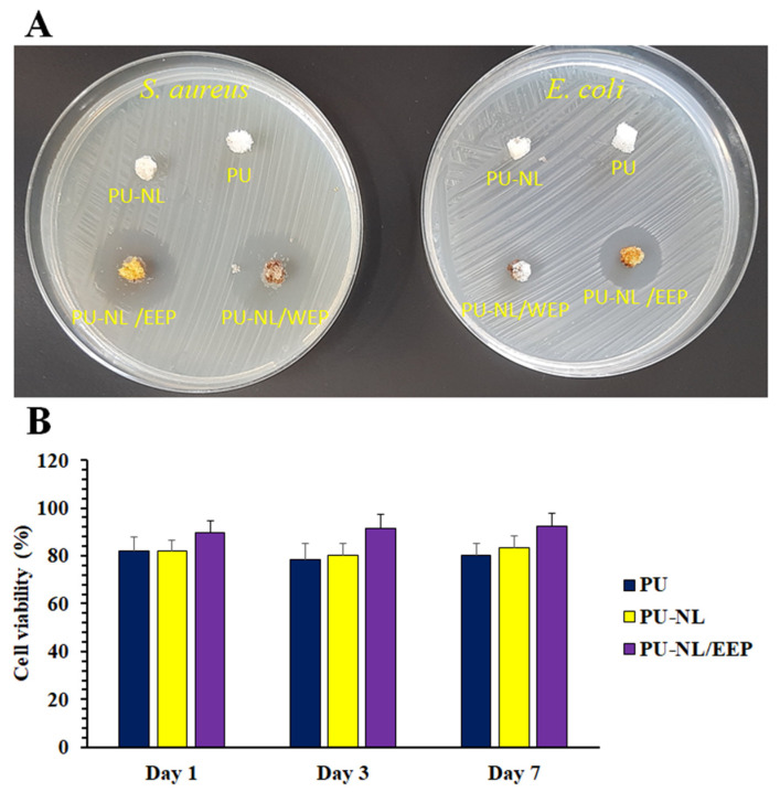
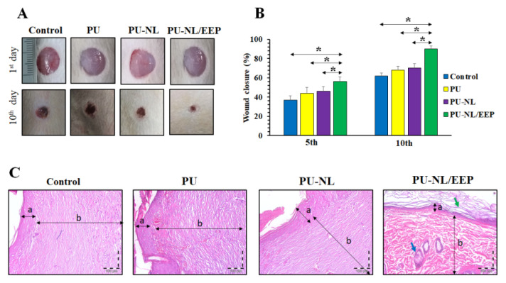
This piece of research explores porous nanocomposite polyurethane (PU) foam synthesis, containing nanolignin (NL), coated with natural antimicrobial propolis for wound dressing. PU foam was synthesized using polyethylene glycol, glycerol, NL, and 1, 6-diisocyanato-hexane (NCO/OH ratio: 1.2) and water as blowing agent. The resultant foam was immersed in ethanolic extract of propolis (EEP). PU, NL-PU, and PU-NL/EEP foams were characterized from mechanical, morphological, and chemical perspectives. NL Incorporation into PU increased mechanical strength, while EEP coating showed lower strength than PU-NL/EEP. Morphological investigations confirmed an open-celled structure with a pore diameter of 150-200 μm, a density of nearly 0.2 g/cm3,, and porosity greater than 85%, which led to significantly high water absorption (267% for PU-NL/EEP). The hydrophilic nature of foams, measured by the contact angle, proved to be increased by NL addition and EEP coating. PU and PU-NL did not show important antibacterial features, while EEP coating resulted in a significant antibacterial efficiency. All foams revealed high biocompatibility toward L929 fibroblasts, with the highest cell viability and cell attachment for PU-NL/EEP. In vivo wound healing using Wistar rats' full-thickness skin wound model confirmed that PU-NL/EEP exhibited an essentially higher wound healing efficacy compared with other foams. Hence, PU-NL/EEP foam could be a promising wound dressing candidate.
Keywords: nanolignin; polyurethane foam; propolis; wound dressing.
Conflict of interest statement
The authors declare no conflict of interest.
Figures


References
-
- Nakielski P., Pawłowska S., Rinoldi C., Ziai Y., De Sio L., Urbanek O., Zembrzycki K., Pruchniewski M., Lanzi M., Salatelli E., et al. Multifunctional platform based on electrospun nanofibers and plasmonic hydrogel: A smart nanostructured pillow for near-infrared light-driven biomedical applications. ACS Appl. Mater. Interfaces. 2020;12:54328–54342. doi: 10.1021/acsami.0c13266. - DOI - PubMed
-
- Ozkaynak M.U., Atalay-Oral C., Tantekin-Ersolmaz S.B., Güner F.S. Polyurethane films for wound dressing applications. Macromol. Symp. 2005;228:177–184. doi: 10.1002/masy.200551016. - DOI
Grants and funding
LinkOut - more resources
Full Text Sources
