. 2021 Aug 24;13(9):1677.
doi: 10.3390/v13091677.

Specificity, Safety, Efficacy of EGFRvIII-Retargeted Oncolytic HSV for Xenotransplanted Human Glioblastoma

Affiliations

Specificity, Safety, Efficacy of EGFRvIII-Retargeted Oncolytic HSV for Xenotransplanted Human Glioblastoma

Irene Appolloni et al. Viruses. .

Abstract

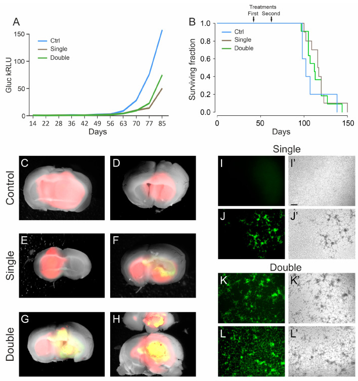
Glioblastoma is a lethal primary brain tumor lacking effective therapy. The secluded onset site, combined with the infiltrative properties of this tumor, require novel targeted therapies. In this scenario, the use of oncolytic viruses retargeted to glioblastoma cells and able to spread across the tumor cells represent an intriguing treatment strategy. Here, we tested the specificity, safety and efficacy of R-613, the first oncolytic HSV fully retargeted to EGFRvIII, a variant of the epidermal growth factor receptor carrying a mutation typically found in glioblastoma. An early treatment with R-613 on orthotopically transplanted EGFRvIII-expressing human glioblastoma significantly increased the median survival time of mice. In this setting, the growth of human glioblastoma xenotransplants was monitored by a secreted luciferase reporter and showed that R-613 is able to substantially delay the development of the tumor masses. When administered as late treatment to a well-established glioblastomas, R-613 appeared to be less effective. Notably the uninfected tumor cells derived from the explanted tumor masses were still susceptible to R-613 infection ex vivo, thus suggesting that multiple treatments could enhance R-613 therapeutic efficacy, making R-613 a promising oncolytic HSV candidate for glioblastoma treatment.

Keywords: EGFRvIII; glioblastoma initiating cells; glioblastoma stem cells; oncolytic HSV; oncolytic virotherapy.


Conflict of interest statement

G.C.-F. is a minor shareholder in Nouscom s.r.l; G.C.-F. and L.M. received equities from Amgen. The funders had no role in the design of the study; in the collection, analyses, or interpretation of data; in the writing of the manuscript, or in the decision to publish the results.

Figures

Figure 1
Figure 1
Specificity and safety of oHSV R-613 infection of glioma cells. (A) Merged fluorescence and bright-field microphotographs of murine glioma cultures expressing EGFRvIII 96 h after infection by R-613 at MOI 1 PFU/cell (I); expressing PDGF-B 72 h after infection by R-613 at MOI 5 PFU/cell (II); expressing PDGF-B 24 h after infection by R-LM5 at MOI 0.2 PFU/cell (wild-type tropism) (III). (B) Merged fluorescence and bright-field microphotographs showing R-613 spread in murine glioma cultures expressing EGFRvIII and infected at MOI 0.3 PFU/cell. (C) Quantification of wtEGFR and EGFRvIII protein expression by Western blot in the indicated glioma cell cultures. (D) Quantification, based on EGFP expression, of the fraction of human glioma cells infected after 24 h by R-613 at the indicated MOI. Dot blots show a representative experiment. (E) Merged fluorescence and bright-field microphotographs showing R-613 spread in EGFRvIII-expressing human glioma cells infected at MOI 0.1 PFU/cell at the indicated time-points. (F,G) Representative dorsal images of brains from NOD/SCID mice treated with R-613 (F) or RLM-5 (G) viruses. Scale bars: 25 μm (A), 50 μm (B,E).
Figure 2
Figure 2
Early treatment with oHSV R-613 on L0306 human glioma cells. (A) Levels of Gluc in blood samples over time in mice transplanted with L0306 human glioma cells infected (blue) or not infected (black) with R-613. Each point represents the moving average of the values with a sliding window of 10 days, centered on the indicated time. In the inset, the same curves are in logarithmic scale. (B) Kaplan–Meyer survival curves of mice transplanted with L0306 human glioma cells infected (blue line, early treatment) or mock infected (black line, control) with R-613. (C) Representative merged fluorescence and brightfield images (dorsal and ventral view) of a mouse brain from the control arm. (D) Representative merged fluorescence and brightfield image of a mouse brain from the early treatment arm and corresponding coronal section at the indicated position (dashed line). (E) Fluorescence and bright-field microphotographs of murine glioma cells expressing EGFRvIII infected with the filtered supernatant of an in vitro culture of a glioma explanted from the early treatment arm. Scale bars: 25 μm.
Figure 3
Figure 3
Late treatment with oHSV R-613 on L0306 human glioma cells. (A) Median Gluc levels over time for each experimental group measured in blood samples collected starting from 14 days after cell transplant, representing in vivo tumor growth (blue line, control; brown line, single treatment; green line, double treatment). (B) Kaplan–Meier survival curves of each experimental arm (same color code as in (A)). (CH) Merged fluorescence and bright-field micrograph images of two representative brains of mice bearing a DsRed human glioma, for each indicated experimental arm. Yellow areas correspond to DsRed-expressing glioma cells infected by EGFP-expressing R-613. (IL) Fluorescence and (I’L’) bright-field microphotographs representing murine glioma cultures expressing human EGFRvIII, inoculated with the filtered supernatant from ex vivo cultured human glioma masses. For each indicated experimental arm, two examples are shown. Scale bars: 25 μm.

References

    1. Bi W.L., Beroukhim R. Beating the Odds: Extreme Long-Term Survival with Glioblastoma. Neuro Oncol. 2014;16:1159–1160. doi: 10.1093/neuonc/nou166. - DOI - PMC - PubMed
    1. Bahadur S., Sahu A.K., Baghel P., Saha S. Current Promising Treatment Strategy for Glioblastoma Multiform: A Review. Oncol. Rev. 2019;13:417. doi: 10.4081/oncol.2019.417. - DOI - PMC - PubMed
    1. Kloos A., Woller N., Gerardy-Schahn R., Kühnel F. Retargeted Oncolytic Viruses Provoke Tumor-Directed T-Cell Responses. Oncoimmunology. 2015;4:e1052933. doi: 10.1080/2162402X.2015.1052933. - DOI - PMC - PubMed
    1. Maroun J., Muñoz-Alía M., Ammayappan A., Schulze A., Peng K.-W., Russell S. Designing and Building Oncolytic Viruses. Future Virol. 2017;12:193–213. doi: 10.2217/fvl-2016-0129. - DOI - PMC - PubMed
    1. Zheng M., Huang J., Tong A., Yang H. Oncolytic Viruses for Cancer Therapy: Barriers and Recent Advances. Mol. Ther. Oncolytics. 2019;15:234–247. doi: 10.1016/j.omto.2019.10.007. - DOI - PMC - PubMed

Publication types

Substances