. 2021 Aug 25;13(9):1685.
doi: 10.3390/v13091685.

The Molecular Tweezer CLR01 Inhibits Antibody-Resistant Cell-to-Cell Spread of Human Cytomegalovirus

Affiliations

The Molecular Tweezer CLR01 Inhibits Antibody-Resistant Cell-to-Cell Spread of Human Cytomegalovirus

Sina Brenner et al. Viruses. .

Abstract

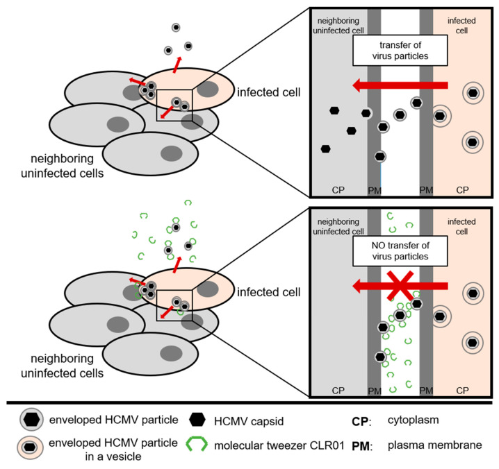
Human cytomegalovirus (HCMV) uses two major ways for virus dissemination: infection by cell-free virus and direct cell-to-cell spread. Neutralizing antibodies can efficiently inhibit infection by cell-free virus but mostly fail to prevent cell-to-cell transmission. Here, we show that the 'molecular tweezer' CLR01, a broad-spectrum antiviral agent, is not only highly active against infection with cell-free virus but most remarkably inhibits antibody-resistant direct cell-to-cell spread of HCMV. The inhibition of cell-to-cell spread by CLR01 was not limited to HCMV but was also shown for the alphaherpesviruses herpes simplex viruses 1 and 2 (HSV-1, -2). CLR01 is a rapid acting small molecule that inhibits HCMV entry at the attachment and penetration steps. Electron microscopy of extracellular virus particles indicated damage of the viral envelope by CLR01, which likely impairs the infectivity of virus particles. The rapid inactivation of viral particles by CLR01, the viral envelope as the main target, and the inhibition of virus entry at different stages are presumably the key to inhibition of cell-free virus infection and cell-to-cell spread by CLR01. Importance: While cell-free spread enables the human cytomegalovirus (HCMV) and other herpesviruses to transmit between hosts, direct cell-to-cell spread is thought to be more relevant for in vivo dissemination within infected tissues. Cell-to-cell spread is resistant to neutralizing antibodies, thus contributing to the maintenance of virus infection and virus dissemination in the presence of an intact immune system. Therefore, it would be therapeutically interesting to target this mode of spread in order to treat severe HCMV infections and to prevent dissemination of virus within the infected host. The molecular tweezer CLR01 exhibits broad-spectrum antiviral activity against a number of enveloped viruses and efficiently blocks antibody-resistant cell-to-cell spread of HCMV, thus representing a novel class of small molecules with promising antiviral activity.

Keywords: CLR01; HCMV; cell-to-cell spread; herpesvirus; inhibition; tweezer.


Conflict of interest statement

The authors declare no conflict of interest.

Figures

Figure 1
Figure 1
CLR01 inhibits HCMV infection. (A,B) Cell-free virus particles of HCMV (strain TB40/E, corresponding to an infection of about 50%) were incubated with indicated concentrations of CLR01 or CLR03 for 30 min at 37 °C prior to infection of human fibroblasts with these mixtures. Infection was determined by indirect immunofluorescence of HCMV IE antigen 24 hpi. (A) Representative images of infected cells: blue, DAPI-positive cells; white, HCMV IE-positive cells. Scale bar is 200 µm. (B) Means ± SDs of relative infection are from the combined results of three individual infection experiments, each performed in triplicate. (C) Cytotoxicity of indicated concentrations of CLR01 and CLR03 on human fibroblasts was controlled after 24 h incubation using the MTT assay. Means ± SDs from two individual experiments, each performed in triplicate. Controls (0 µM) were set to 100% and samples normalized accordingly. (D,E) HCMV-positive urine and saliva (undiluted) were incubated with indicated concentrations of CLR01 for 30 min at 37 °C prior to infection of human fibroblasts with these mixtures for 1 h at 37 °C. Cells were stained for HCMV IE antigen by indirect immunofluorescence at 24 hpi. (D) Means ± SDs of infection (%) and (E) number of HCMV-positive cells per well from triplicate infections. *, p < 0.01; **, p < 0.001; ***, p < 0.0001.
Figure 2
Figure 2
CLR01 reduces the attachment capacity of HCMV particles. (A) Cell-free virus particles of a fluorescent-tagged HCMV were incubated with 50 µM CLR01 and medium for 30 min at 37 °C. Attachment of virus particles to human fibroblasts was allowed for 1 h at 4 °C. Blue, DAPI-stained cell nuclei; yellow, cell-bound HCMV particles; and white, cell borders. Scale bar is 5 µm. (B) Quantification from A with neutralizing antibodies (nAbs, 5 mg/mL Gamunex) as an additional control. Number of attached HCMV particles per cell was determined for at least 100 cells for each condition from two individual experiments. Means ± SDs of relative numbers of attached HCMV particles per cell. Controls (medium) were set to 100% and samples normalized accordingly. ***, p < 0.0001.
Figure 3
Figure 3
CLR01 blocks viral infection post attachment. (A) Cell-free virus particles of HCMV (strain TB40/E) were attached to human fibroblasts for 1 h at 4 °C prior to addition of either 50 µM CLR01 or medium for another hour at 4 °C. Infection was determined at 24 hpi by detection of HCMV IE antigen after the shift to 37 °C. Blue, DAPI-positive cells; white, HCMV-positive cells. Scale bar is 200 µm. (B) Quantification from A with additional controls (neutralizing Abs (nAbs, 5 mg/mL Gamunex), anti-gH Abs (10 µg/mL), human IgG (HCMV negative, 10 µg/mL), elite human serum (HCMV positive, 1:10)), and indicated concentrations of CLR01. Means ± SDs of relative infection are from the combined results of two individual experiments, each performed in triplicate. Controls (medium) were set to 100% and samples normalized accordingly. *, p < 0.01; **, p < 0.001; ***, p < 0.0001.
Figure 4
Figure 4
CLR01 rapidly inactivates HCMV particles. Cell-free virus particles of HCMV (strain TB40/E) were incubated with 50 µM CLR01 for indicated times at 37 °C prior to inactivation of CLR01 by adding medium containing fetal calf serum (FCS; final concentration 20%) and infection of human fibroblasts with these mixtures. Infection was determined after detection of HCMV IE antigen by indirect immunofluorescence at 24 hpi. (A) Blue, DAPI-positive cells; white, HCMV-positive cells. Scale bar is 200 µm. (B) Means ± SDs of relative infection are from the combined results of two individual experiments, each performed in triplicate. Control (0 min) was set to 100% and samples normalized accordingly. ***, p < 0.0001.
Figure 5
Figure 5
CLR01 inhibits direct cell-to-cell spread of a clinical HCMV isolate. (A) Human fibroblasts were seeded together with cells infected with a clinical HCMV isolate. Different conditions (medium, methylcellulose (MC) overlay, HCMV-neutralizing antibodies (nAbs; 5 mg/mL Gamunex), and indicated concentrations of CLR01) were applied 1 day post seeding (dps) and renewed every 48 h. Focal growth was determined by indirect immunofluorescence of HCMV IE antigen at 6 dps. Initial infection was controlled by detection of HCMV-positive cells at 1 dps. (B) Blue, DAPI-positive cells; white, HCMV-positive cells. Scale bar is 200 µm. (C) Means ± SEMs. Each data point represents the number of HCMV-infected cells per focus quantified from images from B. (D) Cell viability of human fibroblasts cultivated under indicated conditions for 6 days at 37 °C was controlled using the MTT assay. Means ± SDs are from the combined results of two individual experiments, each performed in triplicate. Controls (medium) were set to 100% and samples normalized accordingly. ***, p < 0.0001.
Figure 6
Figure 6
CLR01 does not impair HCMV release during infection but alters virion structure. (A) Human fibroblasts were infected with cell-free TB40/E (corresponding to an infection of about 50%) for 1 h and cultured from 1 dpi in either medium or medium containing CLR01. Supernatants were harvested 1, 4, and 6 dpi followed by proteinase K digestion and subsequently used for RT-qPCR. (B) Transmission electron microscopy of extracellular HCMV virions after 6 days of cultivation on human fibroblasts without (left) and with 50 µM CLR01 (right) according to the focus expansion protocol (Supplementary Figure S2). First row: overviews of virions (white arrowheads) in the extracellular space. Scale bar 200 nm. Lower rows: higher magnifications of extracellular virions. Note the circular and regular appearance of virions without CLR01 compared to the altered appearance of virions when treated with CLR01, including the extra layer of electron dense material lining their envelope. Black arrowheads: discontinuities and indentations of the viral envelope. Scale bar: 100 nm.
Figure 7
Figure 7
CLR01 inhibits cell–cell fusion of cells infected with AD169 UL131-repaired HCMV strain. (A) Human fibroblasts were infected with AD169 UL131-repaired HCMV strain (corresponding to an infection of about 0.1–1%). Different conditions (medium, methylcellulose (MC) overlay, HCMV-neutralizing antibodies (nAbs; 5 mg/mL Gamunex), and indicated concentrations of CLR01) were applied 1 dpi and renewed every 48 h. Focal growth and the formation of cell–cell fusion were determined by indirect immunofluorescence of HCMV tegument protein pp65 at 6 dpi. Blue, DAPI-positive cells; green, HCMV-positive cells; white, cell borders. Scale bar is 50 µm. (B) Focus expansion assays were performed with AD169 and AD169 UL131-repaired strain as described in A. Cell–cell fusion events were quantified from triplicate infection. A cell–cell fusion event was defined as at least two cell nuclei within one cell, whereby the different numbers of cell nuclei within cell–cell fusion events was not considered.
Figure 8
Figure 8
CLR01 inhibits cell-to-cell spread of HSV-1 and HSV-2. Human fibroblasts were infected with indicated viruses (corresponding to an infection of about 0.1–1%). Methylcellulose (MC) overlay or 50 µM CLR01 were added at 1 hpi, and the virus spread was examined at 18 hpi. Virus dissemination was determined from the GFP signal of HSV-2-infected cells and from indirect immunofluorescence staining of ICP0 of HSV-1-infected cells. Blue, DAPI-positive cells; green, HSV-positive cells. Scale bar is 200 µm. Means ± SDs of infected area (%) quantified from at least three randomly taken images for each condition. ***, p < 0.0001.
Figure 9
Figure 9
Model of HCMV cell-to-cell spread in human fibroblasts. Upper panel: Unrestricted transfer of enveloped HCMV particles between cell–cell contacts. Lower panel: CLR01 blocks transfer of enveloped HCMV particles between cell–cell contacts.

Similar articles

Cited by

References

    1. Sattentau Q. Avoiding the void: Cell-to-cell spread of human viruses. Nat. Rev. Microbiol. 2008;6:815–826. doi: 10.1038/nrmicro1972. - DOI - PubMed
    1. Krawczyk A., Arndt M.A.E., Grosse-Hovest L., Weichert W., Giebel B., Dittmer U., Hengel H., Jäger D., Schneweis K.E., Eis-Hübinger A.M., et al. Overcoming drug-resistant herpes simplex virus (HSV) infection by a humanized antibody. Proc. Natl. Acad. Sci. USA. 2013;110:6760–6765. doi: 10.1073/pnas.1220019110. - DOI - PMC - PubMed
    1. Clementi N., Criscuolo E., Cappelletti F., Quaranta P., Pistello M., Diotti R.A., Sautto G.A., Tarr A.W., Mailland F., Concas D., et al. Entry inhibition of HSV-1 and -2 protects mice from viral lethal challenge. Antivir. Res. 2017;143:48–61. doi: 10.1016/j.antiviral.2017.03.028. - DOI - PubMed
    1. Mothes W., Sherer N.M., Jin J., Zhong P. Virus Cell-to-Cell Transmission. J. Virol. 2010;84:8360–8368. doi: 10.1128/JVI.00443-10. - DOI - PMC - PubMed
    1. Ganesh L., Leung K., Loré K., Levin R., Panet A., Schwartz O., Koup R.A., Nabel G.J. Infection of specific dendritic cells by CCR5-tropic human immunodeficiency virus type 1 promotes cell-mediated transmission of virus resistant to broadly neutralizing antibodies. J. Virol. 2004;78:11980–11987. doi: 10.1128/JVI.78.21.11980-11987.2004. - DOI - PMC - PubMed

Publication types

MeSH terms

LinkOut - more resources