. 2021 Aug 31;13(9):1733.
doi: 10.3390/v13091733.

Structure-Guided Creation of an Anti-HA Stalk Antibody F11 Derivative That Neutralizes Both F11-Sensitive and -Resistant Influenza A(H1N1)pdm09 Viruses

Affiliations

Structure-Guided Creation of an Anti-HA Stalk Antibody F11 Derivative That Neutralizes Both F11-Sensitive and -Resistant Influenza A(H1N1)pdm09 Viruses

Osamu Kotani et al. Viruses. .

Abstract

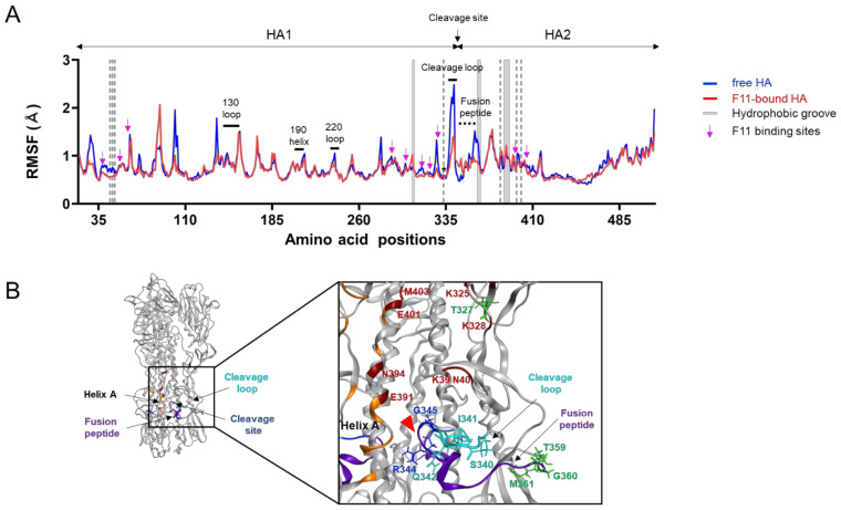
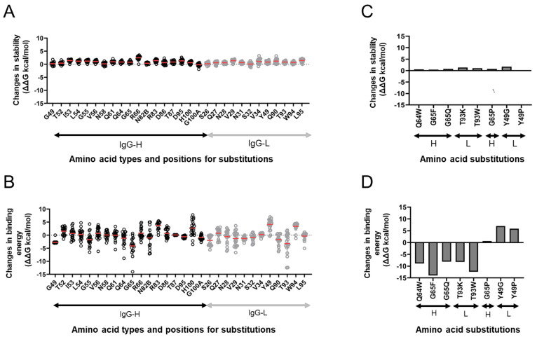
The stalk domain of influenza virus envelope glycoprotein hemagglutinin (HA) constitutes the axis connecting the head and transmembrane domains, and plays pivotal roles in conformational rearrangements of HA for virus infection. Here we characterized molecular interactions between the anti-HA stalk neutralization antibody F11 and influenza A(H1N1)pdm09 HA to understand the structural basis of the actions and modifications of this antibody. In silico structural analyses using a model of the trimeric HA ectodomain indicated that the F11 Fab fragment has physicochemical properties, allowing it to crosslink two HA monomers by binding to a region near the proteolytic cleavage site of the stalk domain. Interestingly, the F11 binding allosterically caused a marked suppression of the structural dynamics of the HA cleavage loop and flanking regions. Structure-guided mutagenesis of the F11 antibody revealed a critical residue in the F11 light chain for the F11-mediated neutralization. Finally, the mutagenesis led to identification of a unique F11 derivative that can neutralize both F11-sensitive and F11-resistant A(H1N1)pdm09 viruses. These results raise the possibility that F11 sterically and physically disturbs proteolytic cleavage of HA for the ordered conformational rearrangements and suggest that in silico guiding experiments can be useful to create anti-HA stalk antibodies with new phenotypes.

Keywords: anti-HA stalk antibody; antibody modification; influenza virus; molecular dynamics simulation; molecular interactions; neutralization assay.


Conflict of interest statement

The authors declare no conflict of interest. The funders had no role in the design of the study; in the collection, analyses, or interpretation of data; in the writing of the manuscript; or in the decision to publish the results.

Figures

Figure 1
Figure 1
Molecular modeling of the complex of glycosylated HA trimer and F11 Fab fragment. A three-dimensional model of the unliganded, glycosylated HA trimer ectodomain of A/Narita/1/2009 (H1N1)pdm09 virus [19] was constructed by the homology modeling method using the Molecular Operating Environment (MOE) (Chemical Computing Group Inc., Montreal, QC, Canada), glycosylated using tools in GLYCAM-Web [21], and subjected to MD simulation using modules in the Amber 16 program package [23]. The HA trimer model in the equilibrium state under solution conditions was used for the docking simulations of the F11 Fab fragment using the Dock application of MOE [29,30]. (A) Root mean square deviation (RMSD) [23] between the structure of the initial HA structure and those at given time points of the MD simulation of glycosylated HA trimers calculated using the cpptraj module in AmberTools 16 as described previously [22]. (B) Side view of the HA trimer model at 400 ns of MD simulation. (C) Distribution of binding energies among the top 100 binding poses generated using the Dock application of MOE [29,30]. (D) Superposition of 100 docking poses. The purple region indicates the HA trimer. Green regions indicate the hydrophobic groove [50] in the HA trimer [53,54]. Grey regions indicate F11 Fab variable regions of the top 100 docking poses. Glycans attached on the HA trimer are not shown here in order to highlight the binding mode of the F11 Fab variable region. (E) The second highest ranked binding mode of F11 Fab (top-2 binding mode). Cyan branches indicate the high-mannose oligosaccharide Man5GlcNAc2.
Figure 2
Figure 2
MD simulation of glycosylated HA trimer docked to the F11 Fab fragment. The top-2 complex was subjected to MD simulation at 1 atm and at 310 K in 0.15 M NaCl for 200 ns. (A) RMSDs between the structure of the initial HA-F11 Fab complex and those at the given time points of the MD simulation are shown. (B) RMSDs of structural units of the F11 Fab fragments during the MD simulations. The entire region (Full), variable region (VR), constant region (CR), and unstructured linker region between VR and CR (linker) are shown for the heavy chain (left panels) and light chain (right panels). (C) Superposition of the top-2 complex structures before and after 200 ns of MD simulations.
Figure 3
Figure 3
Characterization of molecular interactions between the HA trimer ectodomain and F11 Fab fragment. HA-F11 complex structures collected at every 1 ns during 180 to 200 ns after MD simulations of the top-2 binding mode were used to search for the sites supporting molecular interactions between HA and the F11 antibody using the Contact Analysis application of MOE. (A) Visualization of contact sites in the HA trimer (left, wine red residues) and F11 Fab variable region (right, light green and blue green). (B) Interaction networks between HA and F11 Fab variable regions. Non-covalent interactions detected during MD simulation were visualized using Cytoscape software version 3.8.2 [34] as described previously [33]. (C) Distribution of hydrophobic patches in the binding interface of HA and F11. Hydrophobic patches with a minimum area of 50 Å2 for protein-protein interactions were estimated using the Protein Patch Analyzer tool in MOE as described previously [31]. (D) Variation of individual amino acid residues forming noncovalent interactions during MD simulation. Amino acid sequences of the HA protein of the influenza A virus H1 (n = 36,564) and H3 (n = 53,989) were obtained from the GISAID database [42]. Shannon entropy [43] was calculated for individual contact sites detected during MD simulation as described previously [31,41].
Figure 4
Figure 4
Effects of F11 Fab binding on the structural fluctuations of the HA ectodomain. Free and F11 Fab-bound trimeric HA ectodomains were subjected to MD simulations for 200 ns using the Amber 16 program package [23]. RMSF values of the Cα atoms of individual HA residues were calculated using 10,000 snapshots during 180 to 200 ns of MD simulations as described previously [18,31,32]. (A) Distributions of RMSF in HA. Numbers on the horizontal axes indicate positions in the HA of influenza A/Narita/1/2009 (H1N1)pdm09 [19]. Magenta arrows indicate the residues contacting F11 Fab during 180 to 200 ns of MD simulations of the top-2 binding mode. The gray shadows indicate locations of HA residues constituting the hydrophobic groove. Regions involved in sialic acid binding [63,64], such as the 130 loop, 190 helix, and 220 loop, are merged with black bars. (B) Structure around the proteolytic cleavage loop. The cleavage loop, fusion peptide [64], and hydrophobic groove [53,54] are shown in cyan, purple, and orange, respectively. A red arrowhead indicates the cleavage site. Residues that noncovalently interacted with the F11 Fab fragment during MD simulation are marked with a wine red color.
Figure 5
Figure 5
In silico site-directed mutagenesis. F11 Fab residues capable of noncovalently interacting with HA (Figure 3A) and the surrounding residues were individually exchanged for 19 nonself residues, and changes in the stability and binding affinity of F11 Fab were calculated using the F11/HA complex structure obtained at 200 ns of MD simulations as described previously [44,45,46] using the Protein Design application of MOE. (A,C) Effects on the stability of the F11 Fab fragment. (B,D) Effects on the binding affinity of the F11 Fab fragment to the HA ectodomain. (C,D) Results on F11 mutants used for the experimental mutagenesis.
Figure 6
Figure 6
Experimental mutagenesis. Single amino acid substitutions were introduced into the Fab fragment of the F11 IgG1 antibody according to the information in Figure 5C,D. Affinity-purified soluble IgG1 antibodies were used to assess neutralization activity using humanized MDCK cells and 100 TCID50 of influenza virus as described previously [48,49]. Neutralization activity was defined as the reciprocal of the lowest concentration (µg/mL) of antibody at which A630 was >X as calculated using the formula X = (A cell − A virus)/2. (A) SDS PAGE of the purified F11 IgG fractions used for the neutralization assay. (B) Neutralization activity against the F11-sensitive (H1N1)pdm09 virus [18] (left panel), F11-resistant (H1N1)pdm09 variant C1 [18] (middle panel), and F11-weakly resistant (H1N1)pdm09 variant G6 [18] (right panel). NT activity is presented in the scatter plots as the geometric mean, with the geometric standard deviation from four technical replicates (except for F11 WT: n = 10). Y-axis values represent neutralizing titers, calculated as 100/minimum concentration (μg/mL) of antibody that neutralized the virus. Dotted lines represent the detection limit (y = 1; 100 μg/mL). ** p < 0.01 and **** p < 0.0001, comparing F11 mutants with F11 WT (Kruskal–Wallis test, followed by Dunn’s multiple comparison test). For statistical analysis, a provisional minimum NT activity value (y = 0.5; 200 μg/mL) was applied to samples in which NT activity was below the detection limit.

Similar articles

Cited by

References

    1. Altman M.O., Angeletti D., Yewdell J.W. Antibody immunodominance: The key to understanding influenza virus antigenic drift. Viral Immunol. 2018;31:142–149. doi: 10.1089/vim.2017.0129. - DOI - PMC - PubMed
    1. Wu N.C., Wilson I.A. A perspective on the structural and functional constraints for immune evasion: Insights from influenza virus. J. Mol. Biol. 2017;429:2694–2709. doi: 10.1016/j.jmb.2017.06.015. - DOI - PMC - PubMed
    1. Bhatt S., Holmes E.C., Pybus O.G. The genomic rate of molecular adaptation of the human influenza a virus. Mol. Biol. Evol. 2011;28:2443–2451. doi: 10.1093/molbev/msr044. - DOI - PMC - PubMed
    1. Kirkpatrick E., Qiu X., Wilson P.C., Bahl J., Krammer F. The influenza virus hemagglutinin head evolves faster than the stalk domain. Sci. Rep. 2018;8:10432. doi: 10.1038/s41598-018-28706-1. - DOI - PMC - PubMed
    1. Webster R.G., Bean W.J., Gorman O.T., Chambers T.M., Kawaoka Y. Evolution and ecology of influenza a viruses. Microbiol. Rev. 1992;56:152–179. doi: 10.1128/mr.56.1.152-179.1992. - DOI - PMC - PubMed

Publication types

MeSH terms

Substances

LinkOut - more resources