. 2021 Aug 31;13(9):1737.
doi: 10.3390/v13091737.

Structural Analysis of the Menangle Virus P Protein Reveals a Soft Boundary between Ordered and Disordered Regions

Affiliations

Structural Analysis of the Menangle Virus P Protein Reveals a Soft Boundary between Ordered and Disordered Regions

Melissa N Webby et al. Viruses. .

Abstract

The paramyxoviral phosphoprotein (P protein) is the non-catalytic subunit of the viral RNA polymerase, and coordinates many of the molecular interactions required for RNA synthesis. All paramyxoviral P proteins oligomerize via a centrally located coiled-coil that is connected to a downstream binding domain by a dynamic linker. The C-terminal region of the P protein coordinates interactions between the catalytic subunit of the polymerase, and the viral nucleocapsid housing the genomic RNA. The inherent flexibility of the linker is believed to facilitate polymerase translocation. Here we report biophysical and structural characterization of the C-terminal region of the P protein from Menangle virus (MenV), a bat-borne paramyxovirus with zoonotic potential. The MenV P protein is tetrameric but can dissociate into dimers at sub-micromolar protein concentrations. The linker is globally disordered and can be modeled effectively as a worm-like chain. However, NMR analysis suggests very weak local preferences for alpha-helical and extended beta conformation exist within the linker. At the interface between the disordered linker and the structured C-terminal binding domain, a gradual disorder-to-order transition occurs, with X-ray crystallographic analysis revealing a dynamic interfacial structure that wraps the surface of the binding domain.

Keywords: Paramyxoviruses; RNA-dependent RNA polymerase; X-ray crystallography; analytical ultracentrifugation; conformational exchange; intrinsically disordered proteins; pararubulaviruses; protein self-association; small-angle X-ray scattering; solution NMR spectroscopy.


Conflict of interest statement

The authors declare no conflict of interest.

Figures
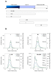
Figure 1
Figure 1
The MenV P coiled-coil drives tetramer formation. (A) A schematic showing the truncated proteins employed in this study; (B) SEC-MALLS data for four individual proteins: P267–328 “L”; P337–388 “BD”; P267–388 ”L-BD”; P209–388 ”CC-L-BD” as indicated. Solid lines show the SEC elution profile (change in refractive index versus elution volume) at the indicated protein loading concentrations. Hollow circles show the mass averaged molar mass estimates resulting from MALLS. Proteins L, BD and L-BD were analyzed using Superdex 75 media (Cytiva), and the plots employ a common vertical scale for refractive index. Protein CC-L-BD was analyzed using Supedex 200 media (Cytiva), and the plot employs a differing vertical scale for refractive index. Molar mass estimates are displayed over the full width at half maximum peak height, in all cases.
Figure 2
Figure 2
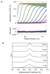
MenV P exists as a tetramer in the micro-molar concentration range. The figure shows sedimentation velocity analytical ultra-centrifugation data and its direct modeling using the program SEDFIT. (A) Time-dependent absorbance scans of CC-L-BD (50 μM concentration) undergoing sedimentation (hollow circles), together with the fit model (solid black lines). Every fourth scan is shown, with the model residuals displayed in the bottom panel. (B) Sedimentation coefficient distributions, c(s), as a function of protein concentration.
Figure 3
Figure 3
A dimer–tetramer equilibrium becomes apparent at low micromolar protein concentrations. The figure shows sedimentation equilibrium analytical ultra-centrifugation data (hollow circles) for CC-L-BD at three different protein concentrations and two different rotor speeds, and its global fit to a rapid reversible dimer–tetramer self-association model (solid lines). Model residuals are shown below the main plots.
Figure 4
Figure 4
Model-free analysis of SAXS data confirms that the linker is globally disordered. The panels on the left show the Kratky plots (I*q2 vs. q) for CC-L-BD (Top), L-BD (middle) and L (bottom). For comparison, the Kratky plot expected of a completely flexible, infinitely thin Gaussian chain, with the same overall radius of gyration (Rg) is shown with a solid red curve. The form factor of the Gaussian chain is given by the Debye function Pq=2x2x1+ex, where x = (q*Rg)2 [78]. The panels on the right show the correspondent pair distance distribution function P(r). The pair distance distribution functions were calculated by indirect Fourier transformation of the experimental SAXS profiles, as implemented in the program GNOM [50]. The radius of gyration, calculated by numerical integration of the pair distribution functions, is indicated.
Figure 5
Figure 5
SAXS data for the linker can be fit to a worm-like-chain (Kratky–Porod) model, providing estimates for the contour length and persistence length. Hollow circles show the SAXS data for the linker on a log-linear scale. The red line shows the fit of the model over the q range 0.015–0.30 Å−1. Best fit model parameters: contour length 192 Å and persistence length 13.5 Å.
Figure 6
Figure 6
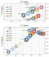
Solution NMR data suggests residues adjacent to the binding domain (residues 325–333) are undergoing temperature-dependent conformational exchange. Selected regions of 2D 1H-15N HSQC spectra, collected at temperatures between 5 and 25 °C, are overlaid in the two panels, with identical iso-contours displayed. As expected [86], the amide chemical shifts track fairly linearly with temperature in all cases. For the residues T325 (bottom panel) and S330 (top panel), which are typical of this region, the backbone amide peak intensities reduce markedly with temperature, with the S330 resonance broadened beyond the detection limit at temperatures >15 °C.
Figure 7
Figure 7
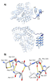
Crystallographic analysis reveals a latch preceding the canonical three-helix bundle of the binding domain. (A) The structure of MenV P329–388 (blue) fused to MBP (grey), in ribbon representation (P212121 crystal form, PDB ID 7KD5). Two orthogonal views are shown. The previously determined structure of the binding domain (residues 339–388) is displayed in light blue, while the newly identified structure (residues 329–338) is displayed in navy blue. (B) Selected residues that mediate formation of the latch and the N-terminal cap of helix α1 are shown in stick representation (P212121 crystal form, PDB ID 7KD5). Hydrogen bonding interactions are shown by the yellow dashed lines. The program UCSF ChimeraX was used to prepare the figures [88].
Figure 8
Figure 8
Crystallographic analysis suggests the latch preceding the binding domain is dynamic. Mean isotropic displacement parameters (B-factors) for main chain atoms are shown for the final helix of MBP and MenV P329–388, which incorporates the last part of the linker and the binding domain. Positions of the alpha helical secondary structures, as defined by the DSSP algorithm [89], are indicated with gray background shading.

References

    1. Drexler J.F., Corman V.M., Müller M.A., Maganga G.D., Vallo P., Binger T., Gloza-Rausch F., Cottontail V.M., Rasche A., Yordanov S., et al. Bats host major mammalian paramyxoviruses. Nat. Commun. 2012;3:796. doi: 10.1038/ncomms1796. - DOI - PMC - PubMed
    1. Smith I., Wang L.-F. Bats and their virome: An important source of emerging viruses capable of infecting humans. Curr. Opin. Virol. 2013;3:84–91. doi: 10.1016/j.coviro.2012.11.006. - DOI - PMC - PubMed
    1. Hauser N., Gushiken A., Narayanan S., Kottilil S., Chua J. Evolution of Nipah Virus Infection: Past, Present, and Future Considerations. Trop. Med. Infect. Dis. 2021;6:24. doi: 10.3390/tropicalmed6010024. - DOI - PMC - PubMed
    1. Clayton B.A. Nipah virus: Transmission of a zoonotic paramyxovirus. Curr. Opin. Virol. 2017;22:97–104. doi: 10.1016/j.coviro.2016.12.003. - DOI - PubMed
    1. Ong K.C., Wong K.T. Henipavirus Encephalitis: Recent Developments and Advances. Brain Pathol. 2015;25:605–613. doi: 10.1111/bpa.12278. - DOI - PMC - PubMed

Publication types

LinkOut - more resources