. 2021 Sep 5;13(9):1771.
doi: 10.3390/v13091771.

Zika Virus Non-Structural Protein 1 Antigen-Capture Immunoassay

Affiliations

Zika Virus Non-Structural Protein 1 Antigen-Capture Immunoassay

Brandon J Beddingfield et al. Viruses. .

Abstract

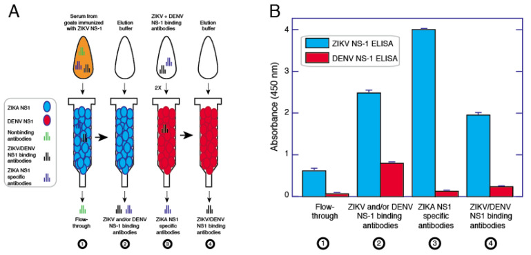
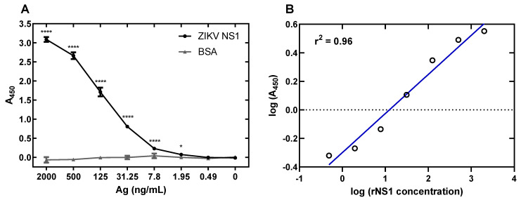
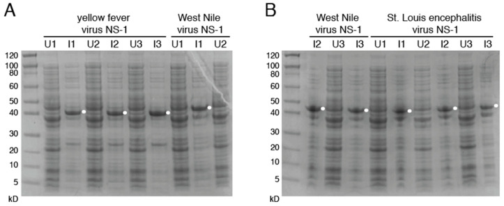
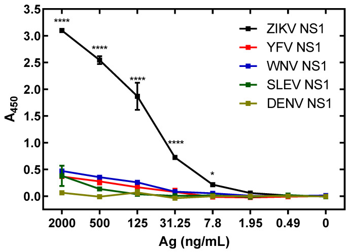
Infection with Zika virus (ZIKV), a member of the Flavivirus genus of the Flaviviridae family, typically results in mild self-limited illness, but severe neurological disease occurs in a limited subset of patients. In contrast, serious outcomes commonly occur in pregnancy that affect the developing fetus, including microcephaly and other major birth defects. The genetic similarity of ZIKV to other widespread flaviviruses, such as dengue virus (DENV), presents a challenge to the development of specific ZIKV diagnostic assays. Nonstructural protein 1 (NS1) is established for use in immunodiagnostic assays for flaviviruses. To address the cross-reactivity of ZIKV NS1 with proteins from other flaviviruses we used site-directed mutagenesis to modify putative epitopes. Goat polyclonal antibodies to variant ZIKV NS1 were affinity-purified to remove antibodies binding to the closely related NS1 protein of DENV. An antigen-capture ELISA configured with the affinity-purified polyclonal antibody showed a linear dynamic range between approximately 500 and 30 ng/mL, with a limit of detection of between 1.95 and 7.8 ng/mL. NS1 proteins from DENV, yellow fever virus, St. Louis encephalitis virus and West Nile virus showed significantly reduced reactivity in the ZIKV antigen-capture ELISA. Refinement of approaches similar to those employed here could lead to development of ZIKV-specific immunoassays suitable for use in areas where infections with related flaviviruses are common.

Keywords: Zika virus; antigen-capture ELISA; non-structural protein 1; polyclonal antibodies; site-directed mutagenesis.


Conflict of interest statement

R.F.G. is a co-founder of Zalgen Labs, a biotechnology company developing countermeasures for emerging viruses. All other authors declare no conflict of interest. The funders had no role in the design of the study; in the collection, analyses, or interpretation of data; in the writing of the manuscript, or in the decision to publish the results.

Figures

Figure 1
Figure 1
Comparison of non-structural protein 1 sequences among closely related flaviviruses. (A): Ribbon model highlighting regions of the NS1 protein containing segments exposed at the outer surface to the host environment. (B): Sequence comparison showing regions with high sequence disparity. Amino acids depicted in red differ from the corresponding ZIKV NS1 amino acids. A represents positions with two sequences with amino acids identical to ZIKV NS1. The boxes highlight highly conserved sequences, amino acids 117–119 and 227–229, that were mutated to alanine in immunodominant regions 2 and 3.
Figure 2
Figure 2
Western blot of NS1 mutants. (A): The blot was probed with anti-His6 antibody targeted toward the protein N-terminus. (B): The blot was probed with anti-ZIKV NS1 monoclonal antibody targeted toward the C-terminus. Uncropped gels are displayed in Figure S1.
Figure 3
Figure 3
Ratios of mutant vs. wild-type binding of NS1 by patient samples. The blue line at 1 indicates equal binding. Numbers over 1 indicate increased binding to the named mutant, while under 1 indicate increased wild-type binding. (A): Ratios of mutant/wild-type binding from IgG in samples show preferential binding to 117–119 mutant NS1. (B): IgM shows preferential binding of mutant NS1 proteins in samples from the Dominican Republic. Colombia = Colombia samples from suspected ZIKV infection. Dom Rep = Dominican Republic samples from suspected ZIKV infection. 117–119 = ZIKV NS1 W117A, G118A, K119A. 227–229 = ZIKV NS1 H227A, T228A, L229A Comparisons were made using Kruskal–Wallis ANOVA. Asterisks represent significant comparisons (**** p < 0.0001; ** p < 0.01). The full dataset is presented in Table S3.
Figure 4
Figure 4
Serum from goats immunized with ZIKV mutants show differential binding to NS1 proteins. Antibody-capture ELISA was performed on a mixture of serum from two goats immunized with ZIKV NS1 protein. Binding of WT ZIKV NS1 showed lower binding to WT (A), both mutants (B,C), as well as DENV2 WT NS1 (D). Data represent samples run in duplicate. Error bars (standard error of the mean) were smaller than the symbols as drawn. The experiment was repeated a total of three times. The legend in panel (D) also applies to panels (AC).
Figure 5
Figure 5
Process for purifying and cross-adsorbing ZIKV NS1 antibodies. ZIKV NS1 antibodies were affinity purified using a gravity flow column. (A): The bound and eluted fraction was put through a column with DENV NS1 twice. The unbound and eluted (non-DENV reactive) fraction is the cross-adsorbed, ZIKV-specific fraction. (B): Antibodies flowing through the column initially bind ZIKV at low signal strength. Following ZIKV NS1 column elution, strength of binding of the solution goes up, but DENV NS1 reactivity is present. Upon cross-adsorption against DENV NS1, specificity of the pAb solution goes up, as well as its binding avidity to ZIKV NS1.
Figure 6
Figure 6
ZIKV NS1 antigen-capture ELISA limit of detection and dynamic range. (A): ZIKV rNS1 shows binding significantly higher than BSA down to 7.8 ng/mL. The limit of detection of the assay is between 7.8 and 1.95 ng/mL NS1. (B): Linear regression of NS1 detection via ELISA demonstrates assay can quantify protein across a range of values. Comparisons were made using two-way ANOVA with a Holm–Sidak multiple comparisons test. Asterisks represent significant comparisons (**** p < 0.0001; * p < 0.05). Error bars represent the standard error of the mean.
Figure 7
Figure 7
Production of flavivirus NS1. Coomassie-stained SDS-PAGE images of induced (I) and uninduced (U) cultures from NS1 proteins of yellow fever virus (A), West Nile virus (A,B) and St. Louis Encephalitis virus (B). Bands indicate proteins similar to DENV and ZIKV NS1 WT production. Results from three different colonies [1,2,3] for each flavivirus NS1 are indicated. Uncropped gels are displayed in Figure S3.
Figure 8
Figure 8
The ZIKV NS1 antigen-capture assay demonstrates low cross-reactivity to related viruses. Recombinant NS1 antigens were produced and assayed. A: Even at low levels of NS1 detection, the assay is specific to ZIKV NS1. Comparisons made using two-way ANOVA with a Holm–Sidak multiple comparisons test. Asterisks represent significant comparisons between ZIKV NS1 and all other NS1 proteins (**** p < 0.0001; * p < 0.05). Error bars represent the standard error of the mean.

References

    1. Dick G.W.A., Kitchen S.F., Haddow A.J. Zika virus. I. Isolations and serological specificity. Trans. R. Soc. Trop. Med. Hyg. 1952;46:509–520. doi: 10.1016/0035-9203(52)90042-4. - DOI - PubMed
    1. Dick G.W.A., Haddow A.J. Uganda S virus: A hitherto unrecorded virus isolated from mosquitoes in Uganda. I. Isolation and pathogenicity. Trans. R. Soc. Trop. Med. Hyg. 1952;46:600–618. doi: 10.1016/0035-9203(52)90021-7. - DOI - PubMed
    1. Smithburen K.C., Kerr J.A., Gatne P.B. Neutralizing antibodies against certain viruses in the sera of residents of India. J. Immunol. 1954;72:248–257. - PubMed
    1. Simpson D.I.H. Zika virus infection in man. Trans. R. Soc. Trop. Med. Hyg. 1964;58:335–338. doi: 10.1016/0035-9203(64)90201-9. - DOI - PubMed
    1. Wikan N., Smith D.R. Zika virus: History of a newly emerging arbovirus. Lancet Infect. Dis. 2016;16:e119–e126. doi: 10.1016/S1473-3099(16)30010-X. - DOI - PubMed

Publication types

MeSH terms