

Nano-Hydroxyapatite vs. Xenografts: Synthesis, Characterization, and In Vitro Behavior
- PMID: 34578603
- PMCID: PMC8469747
- DOI: 10.3390/nano11092289
Nano-Hydroxyapatite vs. Xenografts: Synthesis, Characterization, and In Vitro Behavior
Abstract
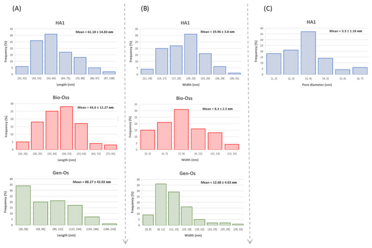
This research focused on the synthesis of apatite, starting from a natural biogenic calcium source (egg-shells) and its chemical and morpho-structural characterization in comparison with two commercial xenografts used as a bone substitute in dentistry. The synthesis route for the hydroxyapatite powder was the microwave-assisted hydrothermal technique, starting from annealed egg-shells as the precursor for lime and di-base ammonium phosphate as the phosphate precursor. The powders were characterized by Fourier-transform infrared spectroscopy (FTIR), X-ray diffraction (XRD), scanning electron microscopy (SEM), energy-dispersive X-ray analysis (EDAX), transmission electron microscopy (TEM), X-ray fluorescence spectroscopy (XRF), and cytotoxicity assay in contact with amniotic fluid stem cell (AFSC) cultures. Compositional and structural similarities or differences between the powder synthesized from egg-shells (HA1) and the two commercial xenograft powders-Bio-Oss®, totally deproteinized cortical bovine bone, and Gen-Os®, partially deproteinized porcine bone-were revealed. The HA1 specimen presented a single mineral phase as polycrystalline apatite with a high crystallinity (Xc 0.92), a crystallite size of 43.73 nm, preferential growth under the c axes (002) direction, where it mineralizes in bone, a nano-rod particle morphology, and average lengths up to 77.29 nm and diameters up to 21.74 nm. The surface of the HA1 nanoparticles and internal mesopores (mean size of 3.3 ± 1.6 nm), acquired from high-pressure hydrothermal maturation, along with the precursor's nature, could be responsible for the improved biocompatibility, biomolecule adhesion, and osteoconductive abilities in bone substitute applications. The cytotoxicity assay showed a better AFSC cell viability for HA1 powder than the commercial xenografts did, similar oxidative stress to the control sample, and improved results compared with Gen-Os. The presented preliminary biocompatibility results are promising for bone tissue regeneration applications of HA1, and the study will continue with further tests on osteoblast differentiation and mineralization.
Keywords: apatite; biomaterial; bone substitute; microwave-assisted hydrothermal synthesis.
Conflict of interest statement
The authors declare no conflict of interest. The funders had no role in the design of the study; in the collection, analyses, or interpretation of data; in the writing of the manuscript, or in the decision to publish the results.
Figures


References
-
- Mello B.F., de Carvalho Formiga M., de Souza da Silva L.F., Dos Santos Coura G., Shibli J.A. Horizontal ridge augmentation using a xenograft bone substitute for implant-supported fixed rehabilitation: A case report with four years of follow-up. Case Rep. Dent. 2020;2020:6723936. doi: 10.1155/2020/6723936. - DOI - PMC - PubMed
-
- Ionescu O.A., Ciocilteu M., Manda V., Neacsu I., Ficai A., Amzoiu E.M., Turcu-Stiolica A., Croitoru O., Neamtu J.O. Bone—Graft delivery systems of type PLGA-gentamicin and Collagen—hydroxyapatite—gentamicine. [(accessed on 15 May 2021)];Mater. Plast. 2019 56:534–537. doi: 10.37358/MP.19.3.5224. Available online: http://www.revmaterialeplastice.ro. - DOI
Grants and funding
- PN-IIIP1-1.2-PCCD-I2017-0629/Unitatea Executiva pentru Finantarea Invatamantului Superior, a Cercetarii, Dezvoltarii si Inovarii
- Project no. 638/12.03.2014, ID 1970/Unitatea Executiva pentru Finantarea Invatamantului Superior, a Cercetarii, Dezvoltarii si Inovarii
- 36355/23.05.2019 HRD OP /380/6/13 - SMIS Code: 123847/European Social Fund
LinkOut - more resources
Full Text Sources
Research Materials
