. 2021 Oct 26;12(5):e0209821.
doi: 10.1128/mBio.02098-21. Epub 2021 Sep 28.

The Burkholderia cenocepacia Type VI Secretion System Effector TecA Is a Virulence Factor in Mouse Models of Lung Infection

Affiliations

The Burkholderia cenocepacia Type VI Secretion System Effector TecA Is a Virulence Factor in Mouse Models of Lung Infection

Nicole A Loeven et al. mBio. .

Abstract

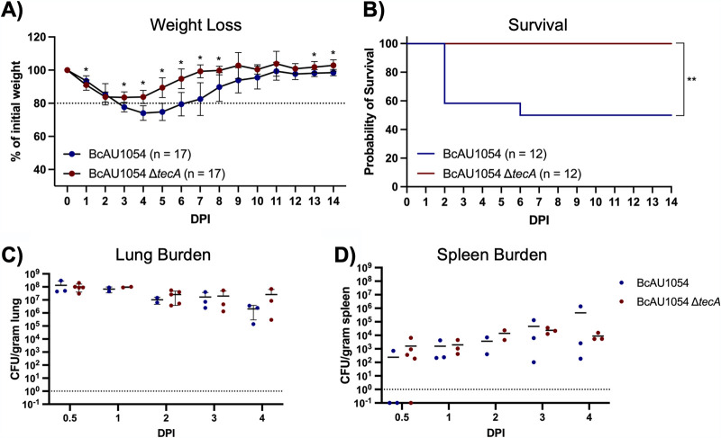
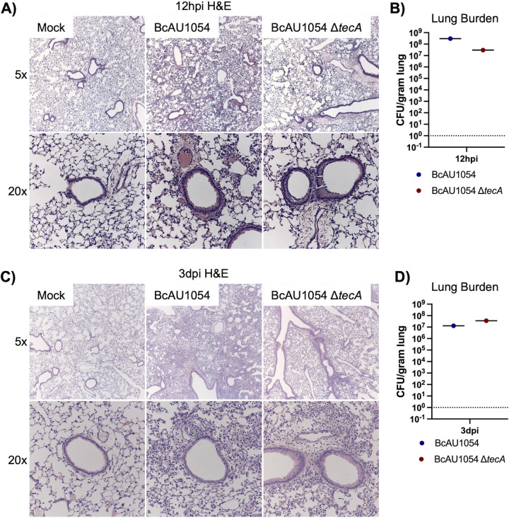
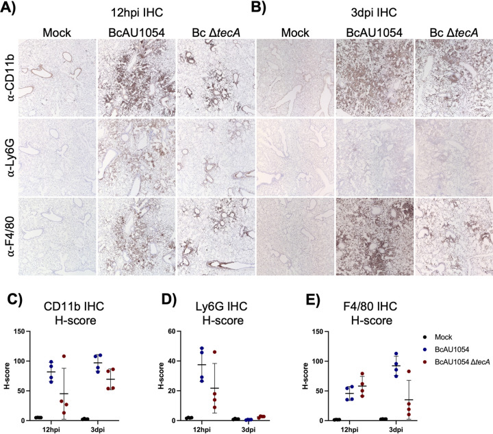
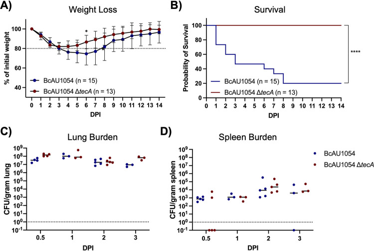
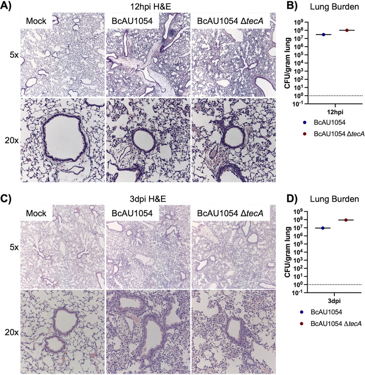
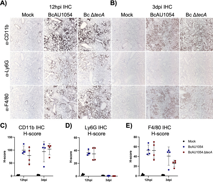
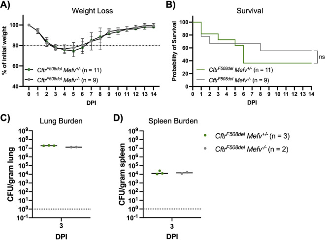
Burkholderia cenocepacia is a member of the Burkholderia cepacia complex (Bcc), a group of bacteria with members responsible for causing lung infections in cystic fibrosis (CF) patients. The most severe outcome of Bcc infection in CF patients is cepacia syndrome, a disease characterized by necrotizing pneumonia with bacteremia and sepsis. B. cenocepacia is strongly associated with cepacia syndrome, making it one of the most virulent members of the Bcc. Mechanisms underlying the pathogenesis of B. cenocepacia in lung infections and cepacia syndrome remain to be uncovered. B. cenocepacia is primarily an intracellular pathogen and encodes the type VI secretion system (T6SS) effector TecA, which is translocated into host phagocytes. TecA is a deamidase that inactivates multiple Rho GTPases, including RhoA. Inactivation of RhoA by TecA triggers assembly of the pyrin inflammasome, leading to secretion of proinflammatory cytokines, such as interleukin-1β, from macrophages. Previous work with the B. cenocepacia clinical isolate J2315 showed that TecA increases immunopathology during acute lung infection in C57BL/6 mice and suggested that this effector acts as a virulence factor by triggering assembly of the pyrin inflammasome. Here, we extend these results using a second B. cenocepacia clinical isolate, AU1054, to demonstrate that TecA exacerbates weight loss and lethality during lung infection in C57BL/6 mice and mice engineered to have a CF genotype. Unexpectedly, pyrin was dispensable for TecA virulence activity in both mouse infection models. Our findings establish that TecA is a B. cenocepacia virulence factor that exacerbates lung inflammation, weight loss, and lethality in mouse infection models. IMPORTANCE B. cenocepacia is often considered the most virulent species in the Bcc because of its close association with cepacia syndrome in addition to its capacity to cause chronic lung infections in CF patients (1). Prior to the current study, virulence factors of B. cenocepacia important for causing lethal disease had not been identified in a CF animal model of lung infection. Results of this study describe a CF mouse model and its use in demonstrating that the T6SS effector TecA of B. cenocepacia exacerbates inflammatory cell recruitment and weight loss and is required for lethality and, thus, acts as a key virulence factor during lung infection. This model will be important in further studies to better understand TecA's role as a virulence factor and in investigating ways to prevent or treat B. cenocepacia infections in CF patients. Additionally, TecA may be the founding member of a family of virulence factors in opportunistic pathogens.

Keywords: Burkholderia cenocepacia; lung infection; type VI secretion.


Figures

FIG 1
FIG 1
TecA enhances weight loss and lethality in WT mice infected with BcAU1054. (A) Weight loss of WT mice infected by o.p. instillation with 5 × 107 CFU of BcAU1054 or ΔtecA mutant. Mice were weighed on D0 and daily for 14 dpi. Results were pooled from 3 independent experiments. n, total mice infected. Data are represented as percentages of initial weight. Error bars are standard deviations. Days on which the two groups differed significantly (P <  0.05, Welch’s t test) are marked with an asterisk. (B) Percent survival monitored for 14 dpi of WT mice infected with BcAU1054 or ΔtecA mutant. Results were pooled from 3 independent experiments. n, total mice infected. **, P = 0.0054 by log-rank (Mantel-Cox) test. (C and D) Lung (C) and spleen (D) burdens at the indicated dpi in WT mice infected with BcAU1054 or ΔtecA mutant. Results are pooled from multiple experiments, and each point represents a value obtained from an individual mouse and data are represented as number of CFU per gram of tissue. Lines are the medians.
FIG 2
FIG 2
Lung inflammation in WT mice infected with BcAU1054 or ΔtecA mutant. (A and C) Representative H&E images of the four right lung lobe sections from a WT mouse left uninfected (mock) or infected o.p. with 5 × 107 CFU of BcAU1054 or ΔtecA mutant at 12 hpi (A) and 3 dpi (C). Light microscopy images are shown at ×5 and ×20 magnification. Burdens of BcAU1054 or ΔtecA mutant in the infected left lungs of the mice analyzed in panels A and C are shown in panels B and D, respectively. Data are represented as number of CFU per gram of lung.
FIG 3
FIG 3
TecA enhances recruitment of neutrophils and inflammatory monocyte-derived macrophages to lungs in WT mice infected with BcAU1054. IHC was performed on sections from the right lung lobes analyzed in Fig. 2 using CD11b, Ly6G, and F4/80 antibodies as indicated. Representative IHC images captured by light microscopy at ×5 magnification are shown. Quantitative IHC image analysis results normalized using H-scores are shown for CD11b (C), for Ly6G (D), and for F4/80 (E). Each data point represents the H-score for each of the four different lobe sections. Error bars are standard deviations, and lines are means.
FIG 4
FIG 4
TecA enhances weight loss and lethality in CftrF508del mice infected with BcAU1054. (A) Weight loss of CftrF508del mice infected o.p. with 5 × 107 CFU of BcAU1054 or ΔtecA mutant. Mice were weighed on D0 and daily for 14 dpi. Results were pooled from 5 independent experiments. n, total mice infected. Data are represented as percentages of initial weight. Error bars are standard deviations. Days on which the two groups differed significantly (P <  0.05, Welch’s t test) are marked with an asterisk. (B) Percent survival monitored for 14 dpi of CftrF508del mice infected with BcAU1054 or ΔtecA mutant. Results were pooled from five independent experiments. n, total mice infected. ****, P < 0.0001 by log-rank (Mantel-Cox) test. (C and D) Lung (C) and spleen (D) burden at the indicated dpi in CftrF508del mice infected with BcAU1054 or ΔtecA mutant. Results were pooled from multiple experiments, and each point represents a value obtained from an individual mouse. Data are represented as number of CFU per gram of tissue. Lines are the medians.
FIG 5
FIG 5
Lung inflammation in CftrF508del mice infected with BcAU1054 or ΔtecA mutant. (A) Representative H&E images of the four right lung lobe sections from a CftrF508del mouse left uninfected (mock) or infected o.p. with 5 × 107 CFU of BcAU1054 or ΔtecA mutant at 12 hpi (A) or 3 dpi (C). Light microscopy images are shown at ×5 and ×20 magnification. Burdens of BcAU1054 or ΔtecA mutant in the infected left lungs of the mice analyzed in panels A and C are shown in panels B and D, respectively. Data are represented as number of CFU per gram of lung.
FIG 6
FIG 6
TecA enhances recruitment of inflammatory monocyte-derived macrophages to lungs in CftrF508del mice infected with BcAU1054. IHC was performed on sections from the right lung lobes analyzed in Fig. 5 using CD11b, Ly6G, and F4/80 antibodies as indicated. Representative IHC images captured by light microscopy at ×5 magnification are shown. Quantitative IHC image analysis results normalized using H-scores are shown for CD11b (C), Ly6G (D), and F4/80 (E). Each data point represents the H-score for each of the four different lobe sections. Error bars are standard deviations, and lines are means.
FIG 7
FIG 7
Pyrin is dispensable for BcAU1054 virulence in CftrF508del mice. (A) Weight loss of CftrF508del Mefv+/ and CftrF508del Mefv−/− mice infected o.p. with 5 × 107 CFU of BcAU1054. Mice were weighed on D0 and daily for 14 dpi. Results were pooled from 3 independent experiments. n, total mice infected. Data are represented as percent initial weight. Error bars are standard deviations. (B) Percent survival monitored for 14 days postinfection of CftrF508del Mefv+/− and CftrF508del Mefv−/− mice infected with BcAU1054. Results were pooled from three independent experiments. n, total mice infected. ns, not significant by log-rank (Mantel-Cox) test. (C and D) Lung (C) and spleen (D) burden at 3 dpi in CftrF508del Mefv+/− and CftrF508del Mefv−/− mice infected with BcAU1054. Data are represented as number of CFU per gram of tissue. Lines are the medians.

References

    1. Loutet SA, Valvano MA. 2010. A decade of Burkholderia cenocepacia virulence determinant research. Infect Immun 78:4088–4100. doi:10.1128/IAI.00212-10. - DOI - PMC - PubMed
    1. Mahenthiralingam E, Urban TA, Goldberg JB. 2005. The multifarious, multireplicon Burkholderia cepacia complex. Nat Rev Microbiol 3:144–156. doi:10.1038/nrmicro1085. - DOI - PubMed
    1. Hauser AR, Jain M, Bar-Meir M, McColley SA. 2011. Clinical significance of microbial infection and adaptation in cystic fibrosis. Clin Microbiol Rev 24:29–70. doi:10.1128/CMR.00036-10. - DOI - PMC - PubMed
    1. Ratjen F, Bell SC, Rowe SM, Goss CH, Quittner AL, Bush A. 2015. Cystic fibrosis. Nat Rev Dis Primers 1:15010. doi:10.1038/nrdp.2015.10. - DOI - PMC - PubMed
    1. Wang XR, Li C. 2014. Decoding F508del misfolding in cystic fibrosis. Biomolecules 4:498–509. doi:10.3390/biom4020498. - DOI - PMC - PubMed

Publication types

LinkOut - more resources