Review
. 2021 Sep 27;12(1):10.
doi: 10.1186/s13227-021-00180-3.

The Nereid on the rise: Platynereis as a model system

Affiliations
Review

The Nereid on the rise: Platynereis as a model system

B Duygu Özpolat et al. Evodevo. .

Abstract

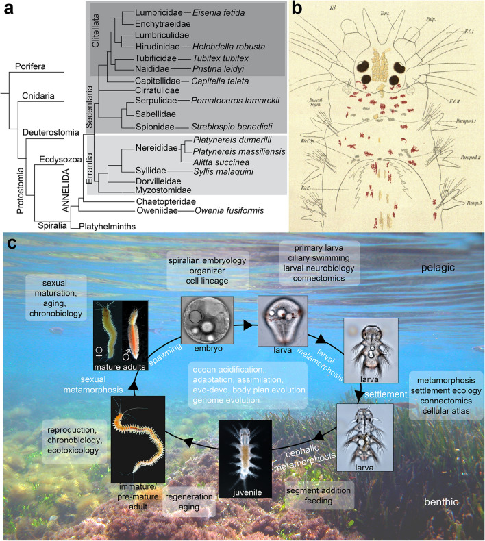
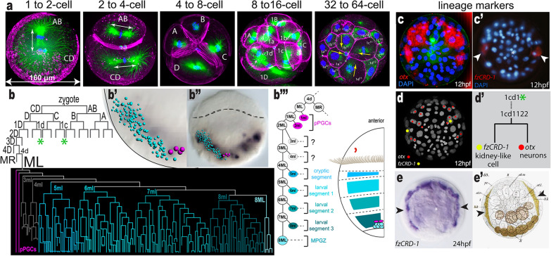
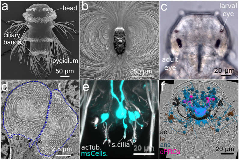
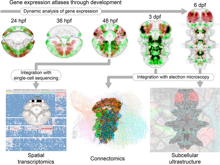
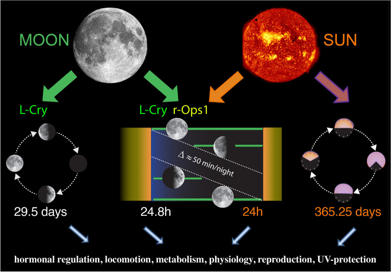
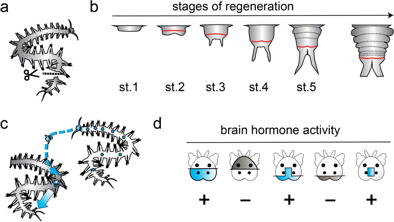
The Nereid Platynereis dumerilii (Audouin and Milne Edwards (Annales des Sciences Naturelles 1:195-269, 1833) is a marine annelid that belongs to the Nereididae, a family of errant polychaete worms. The Nereid shows a pelago-benthic life cycle: as a general characteristic for the superphylum of Lophotrochozoa/Spiralia, it has spirally cleaving embryos developing into swimming trochophore larvae. The larvae then metamorphose into benthic worms living in self-spun tubes on macroalgae. Platynereis is used as a model for genetics, regeneration, reproduction biology, development, evolution, chronobiology, neurobiology, ecology, ecotoxicology, and most recently also for connectomics and single-cell genomics. Research on the Nereid started with studies on eye development and spiralian embryogenesis in the nineteenth and early twentieth centuries. Transitioning into the molecular era, Platynereis research focused on posterior growth and regeneration, neuroendocrinology, circadian and lunar cycles, fertilization, and oocyte maturation. Other work covered segmentation, photoreceptors and other sensory cells, nephridia, and population dynamics. Most recently, the unique advantages of the Nereid young worm for whole-body volume electron microscopy and single-cell sequencing became apparent, enabling the tracing of all neurons in its rope-ladder-like central nervous system, and the construction of multimodal cellular atlases. Here, we provide an overview of current topics and methodologies for P. dumerilii, with the aim of stimulating further interest into our unique model and expanding the active and vibrant Platynereis community.

Keywords: Annelida; Evo-devo; Integrative biology; Marine model species; Spiralia.


Conflict of interest statement

The authors declare that they have no competing interests.

Figures

Fig. 1
Fig. 1
a Phylogenetic position of P. dumerilii in Annelida and in Metazoa [33, 34]. b Platynereis head morphology (from [35]). Ac: acicula; Buccal Segm: buccal segment; F.C. (I, II): tentacular cirri; Kief.: jaw; Palp: palpae; Parapod: parapodia; Tent: antennae. c Life cycle of P. dumerilii. Embryonic, larval, juvenile, and adult stages are shown, along with research areas that focus on the different life cycle stages (boxes). Immature adults: no visible maturing gametes; pre-mature adults: maturing gametes in the coelom; mature adults: fully metamorphosed and ready to spawn
Fig. 2
Fig. 2
Spiral embryogenesis in P. dumerilii: Linking lineage to gene expression and fate cell-by-cell. a Early stages of the P. dumerilii embryo. The mitotic spindles are labeled with anti-alpha-tubulin antibody (green), the actin with rhodamine phalloidin (magenta), and the nucleus with DAPI (blue). Arrows show the direction of cell division. The zygote first divides into AB and CD cells (2-cell stage) and after the second cleavage A, B, C, and D cells are generated. The subsequent spiral cleavages with alternating division axes lead to a 64-cell stage embryo with A, B, C, and D quadrants (described in Hsieh 2020 [70]). bb’’’ P. dumerilii cell lineage with 4d lineage highlighted. Mesoderm (shades of blue), germline (magenta), and mesodermal posterior growth zone (8ML) arise from the 4d lineage. b’’ The segmental mesoderm blocks that arise from this lineage are shown with respect to the larval schematic (b’’’) (modified from Özpolat et al. 2017). ce’ Cell lineage-specific markers (e’ is modified from Wilson 1892, Bastin et al. 2015, Vopalensky et al. 2019) Otx (c,) and fzCRD-1 (c’) expressing cells at 12 hpf were aligned onto the live-imaging movie of a Platynereis embryo used for constructing the anterior cell lineages (d). d’ A subset of the cell lineage from b is shown (*). Otx, which patterns neuronal tissues, and fzCRD-1, which labels a pair of kidney-like cells, are differentially expressed in daughter cells from the same founder cells, 1cd1122. The progeny of the otx-expressing daughter cell will continue to divide and form neurons while the fzCRD-1 expressing daughter cell will cease dividing and differentiate into a kidney-like cell. e At 24 hpf, fzCRD-1 is expressed in two elongated cells that correspond to the pair of kidney-like cells originally observed by E. B. Wilson in his foundational work on P. megalops and Nereis cell lineages in 1892 [ (arrowheads in e’)
Fig. 3
Fig. 3
Locomotor and sensory cilia in the P. dumerilii larva. a SEM micrograph of a larva at 3 days post-fertilization (dpf) (nectochaete stage). Ciliary bands in the head, trunk and tail (pygidium) propel the larva in the water. Dorsal view. b A 3 dpf larva engages in swimming, as visualized by the movement of microbeads added to the water. c Bright-field image of a 3 dpf larva showing the location of adult and larval eyes. Dorsal view. d Transmission electron microscopy image of ciliary photoreceptor cells located in the larval brain. The sensory cilia (outlined in blue) of one of the two pairs of cells is shown. e Mechanosensory ciliated cells (msCells) in the pygidium labeled using a promoter construct. Acetylated tubulin labels sensory cilia (s.cilia). f EM volume reconstruction of the neurosecretory cells in the apical organ region of a 3 dpf larva. Ae: adult eye; le: larval eye; ans: apical nervous system; cPRCs: ciliary photoreceptor cells. Image in a by J. Berger; image in d by R. Shahidi
Fig. 4
Fig. 4
P. dumerilii multimodal atlases. On the top row, five illustrative examples for the ProSPr gene expression atlases at different stages, showing the expression for the genes pax6 in red and gata123 in green. Their co-expression is shown in black. These are sum-projections for the full body from a ventral view (anterior up). On the bottom row, in left–right order: representation of single-cell rna sequencing and spatial mapping to the 48hpf gene expression atlas [61]; reconstructed neurons in the 3dpf P. dumerilii connectome, colored by cell type (the background is a graph representation of such connectome, where nodes represent cells and edges represent synaptic connections) [77, 95]; 2D section of the multimodal cellular atlas for the 6dpf P. dumerilii larva [102] showing the expression of the same genes as the top row, the segmentation of the muscles in cyan, and the segmentation of the chromatin in each nuclei
Fig. 5
Fig. 5
Platynereis dumerilii chronobiology. Sun and moon provide different cues for the rhythms that govern the worms’ biology. L-Cry functions as a valence detector, as well as a detector for the full moon phase. r-Ops1 biochemically cannot discriminate between sun or moonlight, but its high sensitivity allows it to precisely determine moonrise. While the ~ 24-h-long clock relies on the “conventional” circadian transcription factor network, the nature of the circalunar (monthly) clock still remains unknown. It also remains to be tested if the annual changes are just direct responses to environmental changes or controlled by an endogenous annual oscillator
Fig. 6
Fig. 6
Posterior regeneration as a paradigm for studying regenerative mechanisms and their regulation. a Experimental amputation of the posterior part of the body; b key stages (st.1–st.5) of posterior regeneration, re-establishing the growth zone (red), as well as molecular and morphological segment boundaries (dashed lines), reminiscent of the arrangements in posterior growth (right scheme). c Experimental paradigm establishing that transplanted juvenile heads produce a “brain hormone” endowing decerebrated individuals with the capacity to regenerate. d Exemplary results of brain grafts, charting the production of brain hormone (blue) to a medial region of the posterior brain. b Modified after Planques et al. 2019; d modified after Hofmann, 1976
Fig. 7
Fig. 7
A female specimen of Platynereis cf massiliensis in reproduction, with the development of juveniles, from the population of the Vulcano island (Aeolian Archipelago, North Sicily) hydrothermal vent’s system (from Waege et al., 2017, modified)
Fig. 8
Fig. 8
The phylogeographic tree of Platynereis dumerilii and P. massiliensis based on COI analyses (From Waege et al. 2017, modified). Clade 1 and clade 2 represent siblings of P. massiliensis at Ischia (blue dot) and Vulcano (orange dots) vents; clade 3 and clade 4 represent siblings of P. dumerilii, grouped without a clear geographic pattern

References

    1. Ocean Biodiversity Information System. https://obis.org/. Accessed 2 Apr 2021.
    1. Teixeira MAL, Nygren A, Ravara A, Vieira PE, Hernández JC, Costa FO. The small polychaete Platynereis dumerilii revealed as a large species complex with fourteen MOTUs in European marine habitats. Eur Surg. 2021;3(4):e64937.
    1. Wäge J, Valvassori G, Hardege JD, Schulze A, Gambi MC. The sibling polychaetes Platynereis dumerilii and Platynereis massiliensis in the Mediterranean Sea: are phylogeographic patterns related to exposure to ocean acidification? Mar Biol. 2017;164(10):199. doi: 10.1007/s00227-017-3222-x. - DOI
    1. Kisseleva MI. Dynamique et production de la population de Polychète Platynereis dumerilii dans la biocoenose de la Cystoseira en Mer Noire. “Grigore Antipa”. Trav Mus Natl Hist Nat Grigore Antipa. 1971;11:49–58.
    1. Popa LO, Popa OP, Krapal A-M, Iorgu EI, Surugiu V. Fine-scale population genetics analysis of Platynereis dumerilii (Polychaeta, Nereididae) in the Black Sea: how do local marine currents drive geographical differentiation? J Exp Zool A Ecol Genet Physiol. 2014;321(1):41–47. doi: 10.1002/jez.1835. - DOI - PubMed

LinkOut - more resources