Comment
. 2021 Nov 1;27(21):5857-5868.
doi: 10.1158/1078-0432.CCR-19-2384. Epub 2021 Sep 27.

HPV Sequencing Facilitates Ultrasensitive Detection of HPV Circulating Tumor DNA

Affiliations
Comment

HPV Sequencing Facilitates Ultrasensitive Detection of HPV Circulating Tumor DNA

Eric Leung et al. Clin Cancer Res. .

Abstract

Purpose: Human papillomavirus (HPV) DNA offers a convenient circulating tumor DNA (ctDNA) marker for HPV-associated malignancies, but current methods, such as digital PCR (dPCR), provide insufficient accuracy for clinical applications in patients with low disease burden. We asked whether a next-generation sequencing approach, HPV sequencing (HPV-seq), could provide quantitative and qualitative assessment of HPV ctDNA in low disease burden settings.

Experimental design: We conducted preclinical technical validation studies on HPV-seq and applied it retrospectively to a prospective multicenter cohort of patients with locally advanced cervix cancer (NCT02388698) and a cohort of patients with oropharynx cancer. HPV-seq results were compared with dPCR. The primary outcome was progression-free survival (PFS) according to end-of-treatment HPV ctDNA detectability.

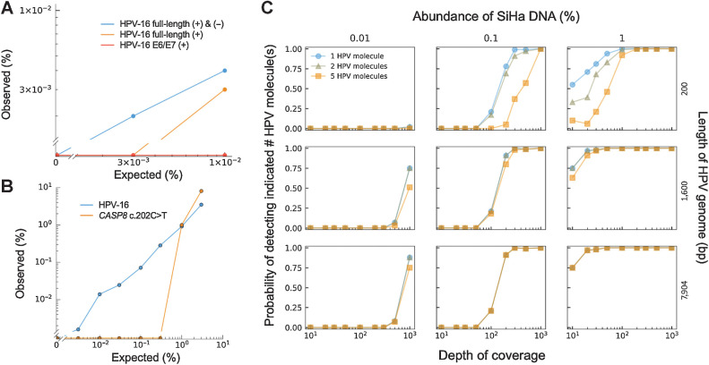
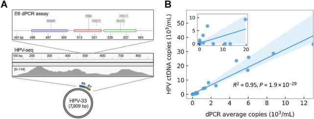
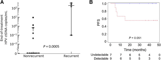
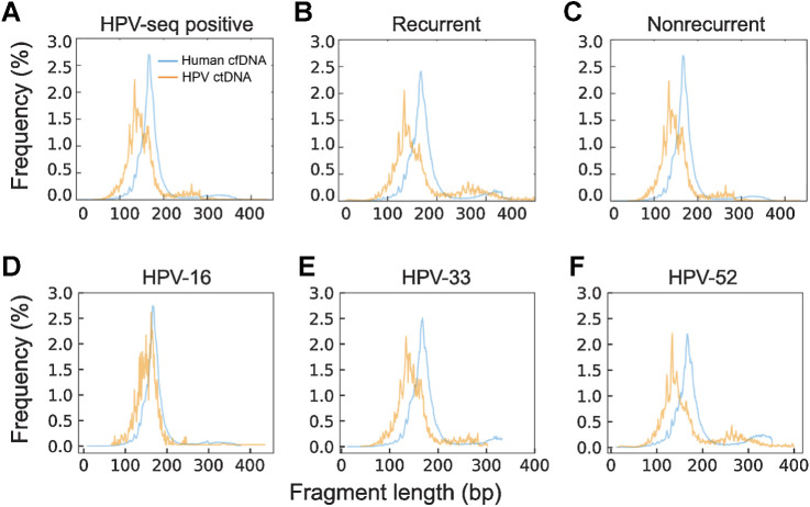
Results: HPV-seq achieved reproducible detection of HPV DNA at levels less than 0.6 copies in cell line data. HPV-seq and dPCR results for patients were highly correlated (R 2 = 0.95, P = 1.9 × 10⁻²⁹) with HPV-seq detecting ctDNA at levels down to 0.03 copies/mL plasma in dPCR-negative posttreatment samples. Detectable HPV ctDNA at end-of-treatment was associated with inferior PFS with 100% sensitivity and 67% specificity for recurrence. Accurate HPV genotyping was successful from 100% of pretreatment samples. HPV ctDNA fragment sizes were consistently shorter than non-cancer-derived cell-free DNA (cfDNA) fragments, and stereotyped cfDNA fragmentomic patterns were observed across HPV genomes.

Conclusions: HPV-seq is a quantitative method for ctDNA detection that outperforms dPCR and reveals qualitative information about ctDNA. Our findings in this proof-of-principle study could have implications for treatment monitoring of disease burden in HPV-related cancers. Future prospective studies are needed to confirm that patients with undetectable HPV ctDNA following chemoradiotherapy have exceptionally high cure rates.


Figures

Figure 1. Overview of HPV-seq and dual-strand hybrid capture. A, HPV-seq conducted on plasma cfDNA is designed to provide quantitative and qualitative information about ctDNA in patients with HPV-associated cancers. In addition to being highly sensitive and quantitative, HPV-seq can report on ctDNA fragment size and HPV genotype. Each full-length viral genome (episome or linearized genome) is expected to yield approximately 50 distinct cfDNA fragments. B, HPV-seq is conducted using hybrid-capture sequencing with single-stranded [sense (+) and/or antisense (−)] biotinylated baits tiled across the HPV genome: (i) single-strand viral genome hybrid capture, (ii) sequential dual-strand hybrid capture, and (iii) simultaneous dual-strand hybrid capture. C, Compared with single-strand hybrid capture (i), dual-strand hybrid capture using either a sequential (ii) or simultaneous (iii) approach recovers more HPV molecules. Results were normalized to the degree of HPV sequence enrichment with a single round of capture using single-stranded baits (left-most bar). Subjecting the unbound library to another round of hybrid capture using baits targeting the same strand (second bar from left) did not improve HPV sequence enrichment. The degree of HPV DNA enrichment in postcapture libraries was determined using HPV-16 E6 and E7 dPCR assays. N = 4 per condition. Error bars represent SD. Asterisk indicates statistical significance (P < 0.05) in comparison with single-strand capture conditions.
Figure 1.
Overview of HPV-seq and dual-strand hybrid capture. A, HPV-seq conducted on plasma cfDNA is designed to provide quantitative and qualitative information about ctDNA in patients with HPV-associated cancers. In addition to being highly sensitive and quantitative, HPV-seq can report on ctDNA fragment size and HPV genotype. Each full-length viral genome (episome or linearized genome) is expected to yield approximately 50 distinct cfDNA fragments. B, HPV-seq is conducted using hybrid-capture sequencing with single-stranded [sense (+) and/or antisense (−)] biotinylated baits tiled across the HPV genome: (i) single-strand viral genome hybrid capture, (ii) sequential dual-strand hybrid capture, and (iii) simultaneous dual-strand hybrid capture. C, Compared with single-strand hybrid capture (i), dual-strand hybrid capture using either a sequential (ii) or simultaneous (iii) approach recovers more HPV molecules. Results were normalized to the degree of HPV sequence enrichment with a single round of capture using single-stranded baits (left-most bar). Subjecting the unbound library to another round of hybrid capture using baits targeting the same strand (second bar from left) did not improve HPV sequence enrichment. The degree of HPV DNA enrichment in postcapture libraries was determined using HPV-16 E6 and E7 dPCR assays. N = 4 per condition. Error bars represent SD. Asterisk indicates statistical significance (P < 0.05) in comparison with single-strand capture conditions.
Figure 2. Analytic sensitivity of HPV-seq. A, HPV-seq was conducted on fragmented SiHa genomic DNA at the indicated dilution. Hybrid capture baits targeted the indicated HPV-16 sequences. The lower limit of detection (LLOD) of HPV-seq was dependent on the use of dual-strand hybrid capture and the length of HPV-16 genome targeted by the baits. B, HPV-seq with full-length dual-strand hybrid capture (blue) provided an improvement in analytic sensitivity and LLOD (0.003%) as compared with hybrid capture for a single mutation (1%). C, Influence of multiple markers and sequencing depth on LLOD. Downsampling of HPV-seq data from full-length dual-strand hybrid capture demonstrates the dependence of the LLOD on the targeted length of HPV-16 genome (i.e., number of markers; right y-axis) and the sequencing depth (x-axis). The probability of detecting the indicated number of HPV molecules (1, blue circles; 2, gray triangles; 5, yellow squares) is shown (left y-axis).
Figure 2.
Analytic sensitivity of HPV-seq. A, HPV-seq was conducted on fragmented SiHa genomic DNA at the indicated dilution. Hybrid capture baits targeted the indicated HPV-16 sequences. The lower limit of detection (LLOD) of HPV-seq was dependent on the use of dual-strand hybrid capture and the length of HPV-16 genome targeted by the baits. B, HPV-seq with full-length dual-strand hybrid capture (blue) provided an improvement in analytic sensitivity and LLOD (0.003%) as compared with hybrid capture for a single mutation (1%). C, Influence of multiple markers and sequencing depth on LLOD. Downsampling of HPV-seq data from full-length dual-strand hybrid capture demonstrates the dependence of the LLOD on the targeted length of HPV-16 genome (i.e., number of markers; right y-axis) and the sequencing depth (x-axis). The probability of detecting the indicated number of HPV molecules (1, blue circles; 2, gray triangles; 5, yellow squares) is shown (left y-axis).
Figure 3. HPV genotyping and ctDNA quantification from plasma cfDNA using HPV-seq. A, HPV-seq correctly genotyped the baseline (pretreatment) plasma cfDNA sample from P19 as HPV-33. HPV-mapping reads were found across the E6 and E7 genes of HPV-33 (100% match to correct genotype). Comparison with dPCR results from the same sample, in which the E6 assay was false negative, reveals mutations in the reverse primer and probe sequences that likely affected dPCR assay performance. B, Comparison of HPV ctDNA copies/mL plasma evaluated by dPCR (x-axis; average of E6 and E7 copies) and HPV-seq (y-axis) for 33 plasma cfDNA samples of patients with cervix cancer obtained at end-of-treatment or posttreatment and 13 plasma cfDNA samples of patients with OPC obtained at pretreatment. Linear regression and its 95% CI (shaded) are shown.
Figure 3.
HPV genotyping and ctDNA quantification from plasma cfDNA using HPV-seq. A, HPV-seq correctly genotyped the baseline (pretreatment) plasma cfDNA sample from P19 as HPV-33. HPV-mapping reads were found across the E6 and E7 genes of HPV-33 (100% match to correct genotype). Comparison with dPCR results from the same sample, in which the E6 assay was false negative, reveals mutations in the reverse primer and probe sequences that likely affected dPCR assay performance. B, Comparison of HPV ctDNA copies/mL plasma evaluated by dPCR (x-axis; average of E6 and E7 copies) and HPV-seq (y-axis) for 33 plasma cfDNA samples of patients with cervix cancer obtained at end-of-treatment or posttreatment and 13 plasma cfDNA samples of patients with OPC obtained at pretreatment. Linear regression and its 95% CI (shaded) are shown.
Figure 4. End-of-treatment detectable HPV ctDNA is associated with disease recurrence. A, HPV ctDNA levels at the end-of-treatment timepoint obtained using HPV-seq are significantly higher among patients who subsequently relapsed (N = 4) versus patients who remained disease-free (N = 12). Horizontal bars indicate the median value and 1.5 times the interquartile range. B, PFS according to HPV ctDNA status at the end-of-treatment timepoint. Data represent HPV copies/mL plasma. Detectable HPV ctDNA, dashed red line. Undetectable HPV ctDNA, solid blue line. Vertical hash marks indicate censoring.
Figure 4.
End-of-treatment detectable HPV ctDNA is associated with disease recurrence. A, HPV ctDNA levels at the end-of-treatment timepoint obtained using HPV-seq are significantly higher among patients who subsequently relapsed (N = 4) versus patients who remained disease-free (N = 12). Horizontal bars indicate the median value and 1.5 times the interquartile range. B, PFS according to HPV ctDNA status at the end-of-treatment timepoint. Data represent HPV copies/mL plasma. Detectable HPV ctDNA, dashed red line. Undetectable HPV ctDNA, solid blue line. Vertical hash marks indicate censoring.
Figure 5. Length distributions of human-mapping cfDNA and HPV-mapping ctDNA fragments for 13 patients with cervix cancer. A, Sequenced fragment insert sizes merged from HPV-seq positive patients analyzed at end-of-treatment or posttreatment (N = 13). Samples (N = 3) from patients with recurrence (B) and samples (N = 10) from patients without recurrence (C) display similar ctDNA fragment-size distributions. Samples from tumors harboring HPV-16 (N = 9; D), HPV-33 (N = 2; E), and HPV-52 (N = 2; F) are shown.
Figure 5.
Length distributions of human-mapping cfDNA and HPV-mapping ctDNA fragments for 13 patients with cervix cancer. A, Sequenced fragment insert sizes merged from HPV-seq positive patients analyzed at end-of-treatment or posttreatment (N = 13). Samples (N = 3) from patients with recurrence (B) and samples (N = 10) from patients without recurrence (C) display similar ctDNA fragment-size distributions. Samples from tumors harboring HPV-16 (N = 9; D), HPV-33 (N = 2; E), and HPV-52 (N = 2; F) are shown.
Figure 6. Genome-wide fragmentomic patterns of HPV-mapping ctDNA fragments across 3 HPV genotypes. First track: Relative read depth coverage for HPV-mapping paired-end reads. Coverage is normalized relative to the maximum for each genome. Relative coverage of 0 indicates no properly paired mapping reads. Samples from tumors harboring HPV-16 (blue; N = 22 samples from 21 patients), HPV-33 (gold; N = 2 samples from 1 patient), and HPV-52 (orange; N = 2 samples from 1 patient) are shown. Second track: Median HPV ctDNA fragment lengths (bp) inferred from sequenced fragment insert sizes. Third track: Capture probes for the 3 HPV genotypes. Fourth track: HPV gene organization from GenBank.
Figure 6.
Genome-wide fragmentomic patterns of HPV-mapping ctDNA fragments across 3 HPV genotypes. First track: Relative read depth coverage for HPV-mapping paired-end reads. Coverage is normalized relative to the maximum for each genome. Relative coverage of 0 indicates no properly paired mapping reads. Samples from tumors harboring HPV-16 (blue; N = 22 samples from 21 patients), HPV-33 (gold; N = 2 samples from 1 patient), and HPV-52 (orange; N = 2 samples from 1 patient) are shown. Second track: Median HPV ctDNA fragment lengths (bp) inferred from sequenced fragment insert sizes. Third track: Capture probes for the 3 HPV genotypes. Fourth track: HPV gene organization from GenBank.

Comment on

  • Selected Articles from This Issue.
    [No authors listed] [No authors listed] Clin Cancer Res. 2021 Nov 1;27(21):5731. doi: 10.1158/1078-0432.CCR-27-21-HI. Clin Cancer Res. 2021. PMID: 34725123 No abstract available.

References

    1. Corcoran RB, Chabner BA. Application of cell-free DNA analysis to cancer treatment. N Engl J Med 2018;379:1754–65. - PubMed
    1. Burgener JM, Rostami A, De Carvalho DD, Bratman SV. Cell-Free DNA as a post-treatment surveillance strategy: Current status. Semin Oncol 2017;44:330–46. - PubMed
    1. Newman AM, Bratman SV, To J, Wynne JF, Eclov NC, Modlin LA, et al. . An ultrasensitive method for quantitating circulating tumor DNA with broad patient coverage. Nat Med 2014;20:548–54. - PMC - PubMed
    1. Shen SY, Singhania R, Fehringer G, Chakravarthy A, Roehrl MHA, Chadwick D, et al. . Sensitive tumour detection and classification using plasma cell-free DNA methylomes. Nature 2018;563:579–83. - PubMed
    1. Abbosh C, Birkbak NJ, Wilson GA, Jamal-Hanjani M, Constantin T, Salari R, et al. . Phylogenetic ctDNA analysis depicts early-stage lung cancer evolution. Nature 2017;545:446–51. - PMC - PubMed

Publication types

Substances