[Preprint]. 2021 Sep 20:rs.3.rs-916427.
doi: 10.21203/rs.3.rs-916427/v1.

Functional antibody and T-cell immunity following SARS-CoV-2 infection, including by variants of concern, in patients with cancer: the CAPTURE study

Annika Fendler  1   2 Lewis Au  1   3   2 Scott T C Shepherd  1   3   2 Fiona Byrne  1 Maddalena Cerrone  4   5 Laura Amanda Boos  3 Karolina Rzeniewicz  1 William Gordon  1 Ben Shum  1   3 Camille L Gerard  1 Barry Ward  1 Wenyi Xie  1 Andreas M Schmitt  3 Nalinie Joharatnam-Hogan  3 Georgina H Cornish  6 Martin Pule  7   8 Leila Mekkaoui  8 Kevin W Ng  6 Eleanor Carlyle  3 Kim Edmonds  3 Lyra Del Rosario  3 Sarah Sarker  3 Karla Lingard  3 Mary Mangwende  3 Lucy Holt  3 Hamid Ahmod  3 Richard Stone  8 Camila Gomes  8 Helen R Flynn  9 Ana Agua-Doce  10 Philip Hobson  10 Simon Caidan  11 Michael Howell  12 Mary Wu  12 Robert Goldstone  13 Margaret Crawford  13 Laura Cubitt  13 Harshil Patel  14 Mike Gavrielides  15 Emma Nye  16 Ambrosius P Snijders  9 James I MacRae  17 Jerome Nicod  13 Firza Gronthoud  18 Robyn L Shea  18   19 Christina Messiou  20 David Cunningham  21 Ian Chau  21 Naureen Starling  21 Nicholas Turner  22 Liam Welsh  23 Nicholas van As  24 Robin L Jones  25 Joanne Droney  26 Susana Banerjee  27 Kate C Tatham  28 Shaman Jhanji  28 Mary O'Brien  29 Olivia Curtis  29 Kevin Harrington  30   31 Shreerang Bhide  30 Jessica Bazin  32 Anna Robinson  32 Clemency Stephenson  32 Tim Slattery  3 Yasir Khan  3 Zayd Tippu  3 Isla Leslie  3 Spyridon Gennatas  33   34 Alicia Okines  22   33 Alison Reid  35 Kate Young  3 Andrew J S Furness  3 Lisa Pickering  3 Sonia Gandhi  36   37 Steve Gamblin  38 Charles Swanton  39   40 Emma Nicholson  32 Sacheen Kumar  21 Nadia Yousaf  29   33 Katalin A Wilkinson  4   41 Anthony Swerdlow  42 Ruth Harvey  43 George Kassiotis  6 James Larkin  3 Robert J Wilkinson  4   5   41 Samra Turajlic  1   3
Affiliations

Functional antibody and T-cell immunity following SARS-CoV-2 infection, including by variants of concern, in patients with cancer: the CAPTURE study

Annika Fendler et al. Res Sq. .

Update in

  • Functional antibody and T cell immunity following SARS-CoV-2 infection, including by variants of concern, in patients with cancer: the CAPTURE study.
    Fendler A, Au L, Shepherd STC, Byrne F, Cerrone M, Boos LA, Rzeniewicz K, Gordon W, Shum B, Gerard CL, Ward B, Xie W, Schmitt AM, Joharatnam-Hogan N, Cornish GH, Pule M, Mekkaoui L, Ng KW, Carlyle E, Edmonds K, Rosario LD, Sarker S, Lingard K, Mangwende M, Holt L, Ahmod H, Stone R, Gomes C, Flynn HR, Agua-Doce A, Hobson P, Caidan S, Howell M, Wu M, Goldstone R, Crawford M, Cubitt L, Patel H, Gavrielides M, Nye E, Snijders AP, MacRae JI, Nicod J, Gronthoud F, Shea RL, Messiou C, Cunningham D, Chau I, Starling N, Turner N, Welsh L, van As N, Jones RL, Droney J, Banerjee S, Tatham KC, Jhanji S, O'Brien M, Curtis O, Harrington K, Bhide S, Bazin J, Robinson A, Stephenson C, Slattery T, Khan Y, Tippu Z, Leslie I, Gennatas S, Okines A, Reid A, Young K, Furness AJS, Pickering L, Gandhi S, Gamblin S, Swanton C; Crick COVID-19 Consortium; Nicholson E, Kumar S, Yousaf N, Wilkinson KA, Swerdlow A, Harvey R, Kassiotis G, Larkin J, Wilkinson RJ, Turajlic S; CAPTURE consortium. Fendler A, et al. Nat Cancer. 2021 Dec;2(12):1321-1337. doi: 10.1038/s43018-021-00275-9. Epub 2021 Oct 27. Nat Cancer. 2021. PMID: 35121900

Abstract

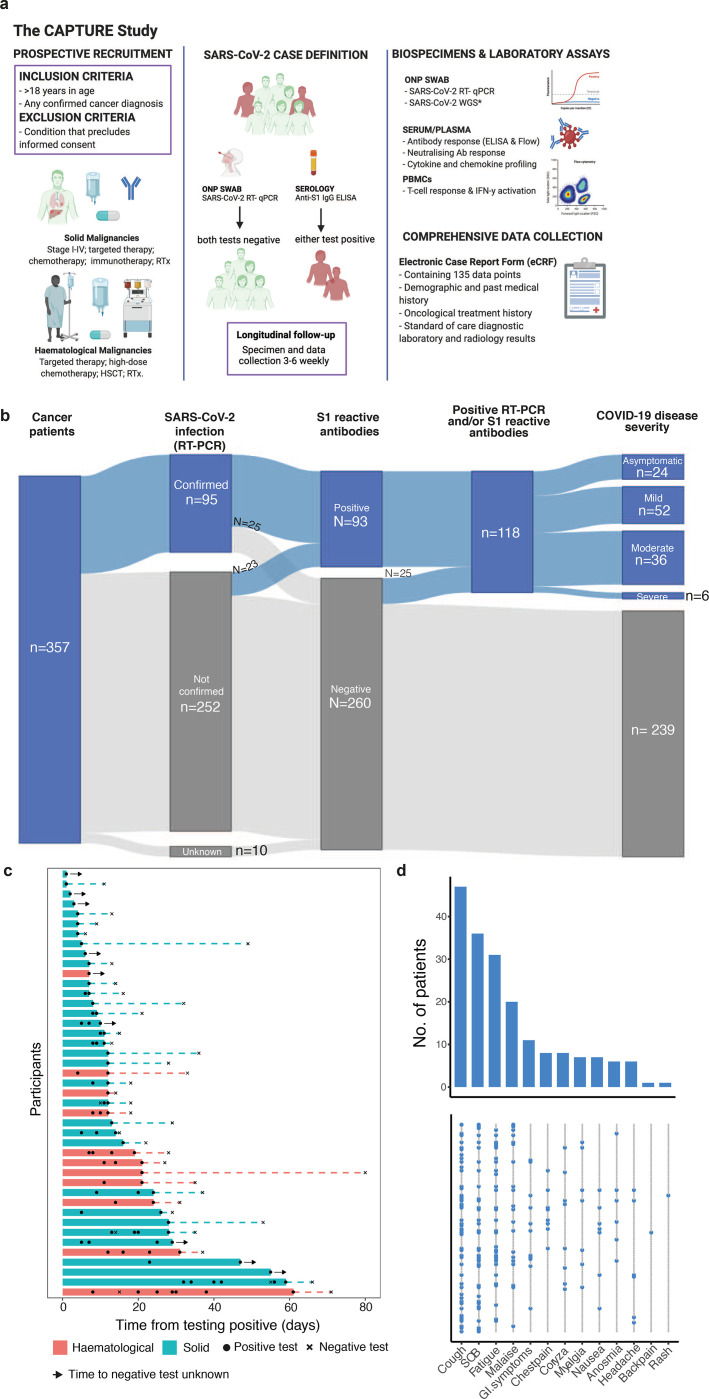
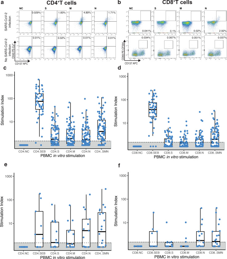
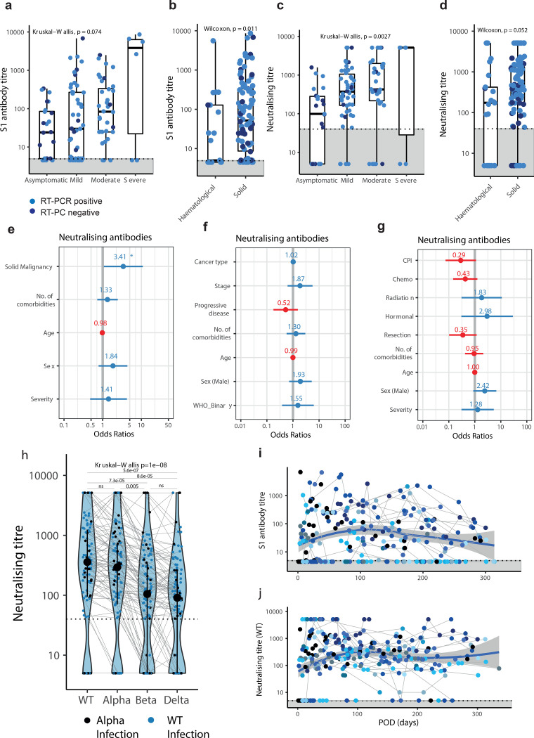
Patients with cancer have higher COVID-19 morbidity and mortality. Here we present the prospective CAPTURE study (NCT03226886) integrating longitudinal immune profiling with clinical annotation. Of 357 patients with cancer, 118 were SARS-CoV-2-positive, 94 were symptomatic and 2 patients died of COVID-19. In this cohort, 83% patients had S1-reactive antibodies, 82% had neutralizing antibodies against WT, whereas neutralizing antibody titers (NAbT) against the Alpha, Beta, and Delta variants were substantially reduced. Whereas S1-reactive antibody levels decreased in 13% of patients, NAbT remained stable up to 329 days. Patients also had detectable SARS-CoV-2-specific T cells and CD4+ responses correlating with S1-reactive antibody levels, although patients with hematological malignancies had impaired immune responses that were disease and treatment-specific, but presented compensatory cellular responses, further supported by clinical. Overall, these findings advance the understanding of the nature and duration of immune response to SARS-CoV-2 in patients with cancer.


Figures

Figure 1:
Figure 1:. SARS-CoV-2 infection status, viral shedding, and COVID-19 symptoms of recruited patients.
a) Patients with cancer irrespective of cancer type, stage, or treatment were recruited. Follow-up schedules for patients with cancer were bespoke to their COVID-19 status and account for their clinical schedules (inpatients: every 2 – 14 days; outpatients: every clinical visit maximum every 3–6 weeks in year one and every six months in year two, and at the start of every or every-second cycle of treatment). Clinical data, oronasopharyngeal swabs and blood were collected at each study visit. Viral antigen testing (RT-PCR on swabs), antibody (ELISA, flow cytometric assay), T cell response and IFN-γ activation assays were performed. b) Distribution of SARS-CoV-2 infection, and S1-reactive Ab status and COVID-19 severity in patients with cancer. 357 patients with cancer were recruited between May 4, 2020 and March 31st 2021. SARS-CoV-2 infection status by RT-PCR and S1-reactive Ab were analysed at recruitment and in serial samples. RT-PCR results prior to recruitment were extracted from electronic patient records. COVID-19 case definition includes all patients with either RT-PCR confirmed SARS-CoV-2 infection or S1-reactive Ab. c) Viral shedding in 43 patients with serial positive swabs. Solid bars indicate time to the last positive test, dotted lines denote the time from the last positive test to the first negative test. d) Distribution of symptoms in 118 COVID-19 patients. Bar graph denotes the number of patients. Each row in the lower graph denotes one patient. ONP, Oronasopharyngeal; ELISA, enzyme-linked immunoassay; PBMCs, peripheral blood mononuclear cells; WGS - whole genome sequencing, RTx, radiotherapy, HSCT, human stem cell transplant.
Figure 2:
Figure 2:. S1-reactive and antibody response in patients with cancer
a) S1-reactive AbT by COVID-19 severity (n=112 patients). Significance was tested by Kruskal-Wallis test, p = 0.074. b) S1-reactive AbT by cancer type (Solid patients: n= 92, Haematological patients: n=20). Significance was tested by two-sided Wilcoxon Wilcoxon-Mann-Whitney U test, p = 0.011. c) NAbT by COVID-19 severity (n=112 patients). Significance was tested by Kruskal-Wallis test, p = 0.0027. d) NAbT by cancer type (Solid patient: n= 92, Haematological patients: n=20). Significance was tested by two-sided Wilcoxon-Mann-Whitney U test, p = 0.052. Boxes indicate 25 and 75 percentiles, line indicates median, and whiskers indicate 1.5 times the IQR. Dots represent individual samples. Dotted lines and grey boxes denote the limit of detection. e) Multivariate binary logistic regression evaluating association with lack of NAb in patients with cancer (n=112). Wald z-statistic was used two calculate two-sided p-values. *, p = 0.038. f) Multivariate binary logistic regression evaluating the association of lack of NAb in patients with solid cancer (n = 92). g) Multivariate binary logistic regression evaluating the association of lack of NAb in patients with solid cancer (n = 92). Dot denotes odds ratio (blue, positive odds ratio; red, negative odds ratio); whiskers indicate 1.5 times the IQR. h) NAbT against WT, Alpha, Beta, and Delta VOCs in patients (n=112) infected with WT SARS-CoV-2 or Alpha VOC. Violin plots denote density of data points. PointRange denotes median and 25 and 75 percentiles. Dots represent individual samples. Significance was tested by Kruskal Wallis test, p = 3.5e-07, two-sided Wilcoxon Mann Whitney U-test with Bonferroni correction (post-hoc test) was used for pairwise comparisons. p-values are denoted in the graph. i) S1-reactive AbT and j) NAbT post onset of disease (n=97 patients). Blue line denotes loess regression line with 95% confidence bands in grey. Black dots denote patients with one sample, coloured dots denote patients with serial samples (n=51 patients). Samples from individual patients are connected. Dotted lines and grey areas at bottom indicate limit of detection. NAb, neutralising antibody, NAbT, neutralising antibody titres, AbT, Antibody titres.
Figure 3:
Figure 3:. T cell response in patients with cancer
a,b) Representative plots of CD4+CD137+OX40+ (CD4+) and CD8+CD137+CD69+ (CD8+) T cells in a patient with confirmed COVID-19 and a cancer patient without COVID-19 after in vitro stimulation with S, M, and N peptide pools, positive control (Staphylococcal enterotoxin B, SEB) or negative control (NC). Frequency of Sars-CoV-2-specific c) CD4+ and d) CD8+ T cells in solid patients with cancer (n= 83). Frequency of Sars-CoV-2-specific e) CD4+ and f) CD8+ T cells in haematological patients with cancer (n= 21). Stimulation index was calculated by dividing the percentage of positive cells in the stimulated sample by the percentage of positive cells in the negative control (NC). To obtain the total number of SsT cells the sum of cells activated by S, M, and N was calculated (SMN). Boxes indicate the 25 and 75 percentiles, line indicates the median, and whiskers indicate 1.5 times the IQR. Individual patients are represented as dots. Dots represent individual samples. Dotted lines and grey boxes denote the limit of detection. SsT cells, Sars-CoV-2-specific T cells.
Figure 4:
Figure 4:. Comparison of antibody and T cell responses in patients with cancer
a) S1-reactive AbT in patients with leukaemia (n=11), myeloma (n=4), and lymphoma (n=6). b) Neutralising antibody titres in patients with leukaemia (n=10), myeloma (n=4), and lymphoma (n=6). c) CD4+ and CD8+ cells T cells across patients with leukemia (n=10), myeloma (n=4), or lymphoma (n=6). Stimulation index was calculated by dividing the percentage of CD4+CD137+OX40+ (CD4+) and CD8+CD137+CD69+ (CD8+) T cells in the stimulated sample by the percentage of positive cells in the negative control (NC). Significance was tested by Kruskal-Wallis test, p < 0.05 was considered significant. d) S1-reactive AbT in patients with haematological malignancy receiving anti-CD20 treatment (n=6) vs other SACT (n=15). e) NAbT in patients with haematological malignancy receiving anti-CD20 treatment (n=6) vs other SACT (n=15). Significance was tested by two-sided Wilcoxon-Mann-Whitney U test, p < 0.05 was considered significant. f) Comparison of CD4+/CD8+ T cells between patients with haematological malignancies on anti-CD20 therapy (n=5, administered within six months) and not on anti-CD20 therapy (n=15). Significance was tested by two-sided Wilcoxon-Mann-Whitney U test, p < 0.05 was considered significant. g) CD4+ and CD8+ cells T cells across patients with solid cancer (n=81) by cancer subtype. Boxes indicate the 25 and 75 percentiles, line indicates the median, and whiskers indicate 1.5 times the IQR. Dots represent individual patient samples. Dotted lines and grey boxes denote the limit of detection. Significance was tested by Kruskal-Wallis test, p < 0.05 was considered significant. SACT, systemic anti-cancer therapy.
Figure 5:
Figure 5:. Associations between SARS-CoV-2-specific T cells with patient or cancer-specific features
Multivariate binary logistic regression analysis evaluating associations between SARS-CoV-2-specific a) CD4+ and b) CD8+ T cells with cancer diagnosis (solid vs haematological malignancies), comorbidities, age, sex, and COVID-19 disease severity in 100 patients. Wald z-statistic was used two calculate two-sided p-values. *, p = 0.038. Multivariate binary logistic regression analysis evaluating associations between SARS-CoV-2-specific c) CD4+ and d) CD8+ T cells with anti-cancer intervention, age, sex, and COVID-19 disease severity in patients with solid cancer (n=81). Wald z-statistic was used two calculate two-sided p-values. *, p = 0.045. Dot denotes odds ratio (blue and red dots indicate positive or negative odds ratio, respectively); whiskers indicate 1.5 times the IQR. e) Comparison of SARS-CoV-2-specific CD4+/CD8+ T cells between patients with solid malignancies on CPI (n=13, administered within three months) and not on CPI (n=68). Boxes indicate the 25th and 75th percentiles, line indicates the median, and whiskers indicate 1.5 times the IQR. Dots represent individual samples. Significance was tested by two-sided Wilcoxon-Mann-Whitney U test (p = 0.038 and 0.53).

References

    1. Williamson E. J. et al.Factors associated with COVID-19-related death using OpenSAFELY. Nature 584, 430–436, doi:10.1038/s41586-020-2521-4 (2020). - DOI - PMC - PubMed
    1. Saini K. S. et al.Mortality in patients with cancer and coronavirus disease 2019: A systematic review and pooled analysis of 52 studies. Eur J Cancer 139, 43–50, doi:10.1016/j.ejca.2020.08.011 (2020). - DOI - PMC - PubMed
    1. Garcia-Suarez J. et al.Impact of hematologic malignancy and type of cancer therapy on COVID-19 severity and mortality: lessons from a large population-based registry study. J Hematol Oncol 13, 133, doi:10.1186/s13045-020-00970-7 (2020). - DOI - PMC - PubMed
    1. Garassino M. C. et al.COVID-19 in patients with thoracic malignancies (TERAVOLT): first results of an international, registry-based, cohort study. Lancet Oncol 21, 914–922, doi:10.1016/S1470-2045(20)30314-4 (2020). - DOI - PMC - PubMed
    1. Lee L. Y. et al.COVID-19 mortality in patients with cancer on chemotherapy or other anticancer treatments: a prospective cohort study. Lancet 395, 1919–1926, doi:10.1016/S0140-6736(20)31173-9 (2020). - DOI - PMC - PubMed

Publication types