. 2021 Nov 19;16(11):2453-2461.
doi: 10.1021/acschembio.1c00553. Epub 2021 Sep 28.

Methylglyoxal Forms Diverse Mercaptomethylimidazole Crosslinks with Thiol and Guanidine Pairs in Endogenous Metabolites and Proteins

Affiliations

Methylglyoxal Forms Diverse Mercaptomethylimidazole Crosslinks with Thiol and Guanidine Pairs in Endogenous Metabolites and Proteins

John S Coukos et al. ACS Chem Biol. .

Abstract

Methylglyoxal (MGO) is a reactive byproduct formed by several metabolic precursors, the most notable being triosephosphates in glycolysis. While many MGO-mediated adducts have been described, the reactivity and specific biomolecular targets of MGO remain incompletely mapped. Based on our recent discovery that MGO can form stable mercaptomethylimidazole crosslinks between cysteine and arginine (MICA) in proteins, we hypothesized that MGO may participate in myriad reactions with biologically relevant guanidines and thiols in proteins, metabolites, and perhaps other biomolecules. Herein, we performed steady-state and kinetic analyses of MGO reactivity with several model thiols, guanidines, and biguanide drugs to establish the plausible and prevalent adducts formed by MGO in proteins, peptides, and abundant cellular metabolites. We identified several novel, stable MICA metabolites that form in vitro and in cells, as well as a novel intermolecular post-translational MICA modification of surface cysteines in proteins. These data confirm that kinetic trapping of free MGO by thiols occurs rapidly and can decrease formation of more stable imidazolone (MG-H1) arginine adducts. However, reversible hemithioacetal adducts can go on to form stable MICA modifications in an inter- and intramolecular fashion with abundant or proximal guanidines, respectively. Finally, we discovered that intracellular MICA-glutathione metabolites are recognized and exported by the efflux pump MRP1, providing a parallel and perhaps complementary pathway for MGO detoxification working alongside the glyoxalase pathway. These data provide new insights into the plausible reactions involving MGO in cells and tissues, as well as several new molecular species in proteins and metabolites for further study.


Conflict of interest statement

The authors declare no competing financial interest.

Figures

Figure 1
Figure 1
Kinetic and thermodynamic characterization of MGO reactions with biological guanidine and thiols. (A) Schematic depicting known and hypothesized MGO reactivity paths with biologically relevant thiols and guanidine-containing metabolites or proteins. (B) Model guanidine- or thiol-containing metabolites used in this study. (C) LC–MS quantification of indicated metabolite reactants, intermediates, and products within time-course studies of equimolar (1 mM) concentrations of the indicated guanidine, thiol, and MGO at 25 °C. (D) LC–MS quantification of indicated reactant and product levels after 24 h incubation of guanidine and thiol compounds (1 mM each) with indicated MGO concentrations at 37 °C. Data plotted in (C,D) are mean with S.E.M. from n = 4 independent biological replicates.
Figure 2
Figure 2
Inter- and intramolecular mercaptomethylimidazole modifications on model peptides and proteins. (A) LC–MS quantification of MGO modifications in the model CRV2 peptide. (B) Logistic fit of MICA formation for peptide time-course experiment and time-course experiments in Figure 1D. (C) LC–MS quantification of MICA product formation under the indicated NAC and Arg cotreatment conditions. (D) Structural depiction (on PDB: 4F5S) of all methylglyoxal-derived modifications observed in BSA treated with MGO and arginine. Chemical structures of each modification are shown (right). (E) Mass spectrum of the Arg-MICA-modified Cys58 peptide detected in BSA. (F) Model summarizing the relative kinetic and thermodynamic landscape of MGO-derived modifications on arginine and cysteine residues. Data plotted in (A–C) represent mean with S.E.M. from independent biological replicates (n = 4).
Figure 3
Figure 3
MICA crosslinks form between endogenous thiol/guanidine metabolite pairs in cells. (A–D) Representative chromatograms of MICA cross-linked metabolites between arginine or metformin and glutathione or cysteine in HeLa cells treated with MGO (blue), metformin (green), both (red), or vehicle (brown) for 8 h along with chromatograms of synthetic standards for the relevant MICA adduct (black). (E–H) Relative LC–MS/MS quantification of arginine, glutathione, MG-H1 arginine, and GSH-Arg-MICA in HeLa cells treated with indicated doses of MGO for 8 h. Relative quantification of each metabolite was based off of the condition in which the maximum MS-transition EIC was detected. Data plotted in (E–H) are mean with S.E.M. (n = 4 independent biological replicates). Statistical analyses are by ordinary one-way analysis of variance (ANOVA). *p < 0.05; **p < 0.01; ***p < 0.001; ****p < 0.0001.
Figure 4
Figure 4
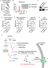
Glutathione-containing MICA metabolites are actively exported by the MRP1 transporter. (A) LC–MS/MS quantification of extracellular GSH-Arg-MICA from HeLa cells treated with indicated doses of MGO for 8 h. (B) Representative chromatograms of intra- and extracellular glutathione levels from HeLa cells treated for 8 h with indicated doses of MGO. (C) Western blot analysis of MRP1 and PGK1 levels in HeLa cells stably transduced with either shABCC1 or control plasmid (scramble). (D–G) LC–MS/MS quantification of intra- and extracellular GSH-Arg-MICA, intracellular arginine, and intracellular glutathione in shABCC1 or scramble HeLa cells treated with 0.5 mM MGO for 8 h. (H) Schematic depicting the deleterious interaction targets of glucose-derived MGO (dashed arrows) and protective capture of MGO by reduced glutathione for glyoxalase-dependent detoxification and, as shown in this work, parallel MICA metabolite formation and excretion from cells. Data plotted in (A) and (D–G) are mean with S.E.M. (n = 4 independent biological replicates). Statistical analysis in (A) is by ordinary ANOVA and (D–G) by unpaired Student’s t-test. *p < 0.05; **p < 0.01; ***p < 0.001; ****p < 0.0001.

References

    1. Alderson N. L.; Wang Y.; Blatnik M.; Frizzell N.; Walla M. D.; Lyons T. J.; Alt N.; Carson J. A.; Nagai R.; Thorpe S. R.; Baynes J. W. S-(2-Succinyl)cysteine: a novel chemical modification of tissue proteins by a Krebs cycle intermediate. Arch. Biochem. Biophys. 2006, 450, 1–8. 10.1016/j.abb.2006.03.005. - DOI - PubMed
    1. Moellering R. E.; Cravatt B. F. Functional lysine modification by an intrinsically reactive primary glycolytic metabolite. Science 2013, 341, 549–553. 10.1126/science.1238327. - DOI - PMC - PubMed
    1. Esterbauer H.; Schaur R. J.; Zollner H. Chemistry and biochemistry of 4-hydroxynonenal, malonaldehyde and related aldehydes. Free Radic. Biol. Med. 1991, 11, 81–128. 10.1016/0891-5849(91)90192-6. - DOI - PubMed
    1. Murphy M. P. How mitochondria produce reactive oxygen species. Biochem. J. 2009, 417, 1–13. 10.1042/bj20081386. - DOI - PMC - PubMed
    1. Chang J. W.; Lee G.; Coukos J. S.; Moellering R. E. Profiling Reactive Metabolites via Chemical Trapping and Targeted Mass Spectrometry. Anal. Chem. 2016, 88, 6658–6661. 10.1021/acs.analchem.6b02009. - DOI - PMC - PubMed

Publication types