Review
. 2021 Nov 1;49(5):2319-2331.
doi: 10.1042/BST20210410.

The pockets guide to HLA class I molecules

Affiliations
Review

The pockets guide to HLA class I molecules

Andrea T Nguyen et al. Biochem Soc Trans. .

Abstract

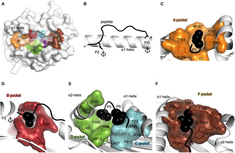
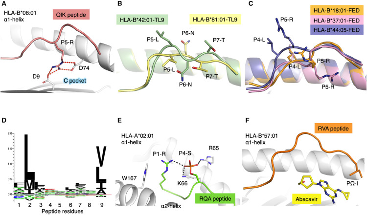
Human leukocyte antigens (HLA) are cell-surface proteins that present peptides to T cells. These peptides are bound within the peptide binding cleft of HLA, and together as a complex, are recognised by T cells using their specialised T cell receptors. Within the cleft, the peptide residue side chains bind into distinct pockets. These pockets ultimately determine the specificity of peptide binding. As HLAs are the most polymorphic molecules in humans, amino acid variants in each binding pocket influences the peptide repertoire that can be presented on the cell surface. Here, we review each of the 6 HLA binding pockets of HLA class I (HLA-I) molecules. The binding specificity of pockets B and F are strong determinants of peptide binding and have been used to classify HLA into supertypes, a useful tool to predict peptide binding to a given HLA. Over the years, peptide binding prediction has also become more reliable by using binding affinity and mass spectrometry data. Crystal structures of peptide-bound HLA molecules provide a means to interrogate the interactions between binding pockets and peptide residue side chains. We find that most of the bound peptides from these structures conform to binding motifs determined from prediction software and examine outliers to learn how these HLAs are stabilised from a structural perspective.

Keywords: HLA; HLA classification; epitope presentation; peptide; peptide binding motif.


Conflict of interest statement

The authors declare that there are no competing interests associated with the manuscript.

Figures

Figure 1.
Figure 1.. HLA pockets representation.
(A) Surface representation of a HLA molecule (white) with the pockets within the cleft shown in different colours. Pocket A (orange), B (red), C (cyan), D (green), E (purple), F (brown) and the peptide shown as a cartoon in white. (B) Peptide represented as a black cartoon with primary anchor residues P2 and PΩ shown as stick against the α1-helix of the HLA shown as a white cartoon. (C) The A pocket of the HLA represented by an orange surface with the residues making up the pocket shown as orange sticks and the surrounding residues represented by a white cartoon. The P1 residue of the peptide is represented by black spheres nested into the A pocket. (D) The B pocket of the HLA represented by a red surface with the residues making up the pocket shown as red sticks and the surrounding residues represented by a white cartoon. The primary anchor P2 residue of the peptide is represented by black spheres. (E) The C pocket of the HLA represented by a cyan surface and residues as sticks, while the D pocket is represented by a green surface with the residues making up the D pocket shown as green sticks. The P3 and P5 of the peptide is represented by black spheres. (F) The F pocket of the HLA represented by a brown surface with the residues making up the pocket shown as brown sticks and the surrounding residues represented by a white cartoon. The PΩ of the peptide is represented by black spheres.
Figure 2.
Figure 2.. HLA polymorphism impacts on peptide presentation and binding.
(A) The α1-helix of HLA-B*08:01 shown as a white cartoon with the residues interacting with the QIK peptide (salmon cartoon) shown as stick. The P5 of the QIK peptide is shown as stick, forming salt bridges (red dashed lines) with the Asp9 and Asp74 of the HLA molecule. (B) Overlay of HLA-B*42:01 (green cartoon) and HLA-B*81:01(yellow cartoon) each presenting the TL9 peptide, in a distinct conformation, shown as cartoon (green and yellow, respectively) with P5, P6 and P7 of the peptide shown as stick. (C) Overlay of HLA-B*18:01(orange cartoon), HLA-B*37:01 (pink cartoon) and HLA-B*44:05 (purple cartoon) presenting the FED peptide shown as cartoon (orange, pink and purple, respectively) with P4 and P5 of each peptide shown as stick. (D) Distribution of preferred peptide residues located into the B and F pockets of HLA-A*02:01 using Seq2logo2.0 [69]. (E) HLA-A*02:01 (white cartoon) with select residues shown as stick presenting the RQA peptide (green cartoon) with P1 shown as stick and P4 represented as an orange stick. The P4-S phosphorylated is represented as stick forming hydrogen bonds (black dashed lines) with the R65 from the HLA and the P1-R from the peptide. (F) HLA-B*57:01 (white cartoon) presenting the RVA peptide (orange cartoon) with the PΩ shown as stick, in the presence of Abacavir in the F pocket of the HLA (yellow stick).

References

    1. Bjorkman, P.J., Saper, M.A., Samraoui, B., Bennett, W.S., Strominger, J.L. and Wiley, D.C. (1987) Structure of the human class I histocompatibility antigen, HLA-A2. Nature 329, 506–512 10.1038/329506a0 - DOI - PubMed
    1. Josephs, T.M., Grant, E.J. and Gras, S. (2017) Molecular challenges imposed by MHC-I restricted long epitopes on T cell immunity. Biol. Chem. 398, 1027–1036 10.1515/hsz-2016-0305 - DOI - PubMed
    1. Jardetzky, T.S., Brown, J.H., Gorga, J.C., Stern, L.J., Urban, R.G., Chi, Y.I.et al. (1994) Three-dimensional structure of a human class II histocompatibility molecule complexed with superantigen. Nature 368, 711–718 10.1038/368711a0 - DOI - PubMed
    1. Sercinoglu, O. and Ozbek, P. (2020) Sequence-structure-function relationships in class I MHC: a local frustration perspective. PLoS ONE 15, e0232849 10.1371/journal.pone.0232849 - DOI - PMC - PubMed
    1. Gfeller, D. and Bassani-Sternberg, M. (2018) Predicting antigen presentation-what could we learn from a million peptides? Front. Immunol. 9, 1716 10.3389/fimmu.2018.01716 - DOI - PMC - PubMed

Publication types

Substances