. 2021 Sep 28;16(9):e0257800.
doi: 10.1371/journal.pone.0257800. eCollection 2021.

Enhanced copper-resistance gene repertoire in Alteromonas macleodii strains isolated from copper-treated marine coatings

Affiliations

Enhanced copper-resistance gene repertoire in Alteromonas macleodii strains isolated from copper-treated marine coatings

Kathleen Cusick et al. PLoS One. .

Abstract

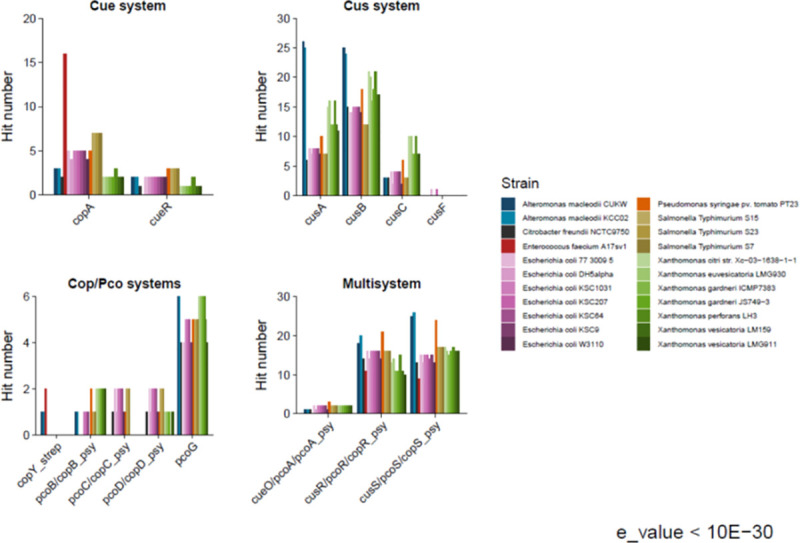
Copper is prevalent in coastal ecosystems due to its use as an algaecide and as an anti-fouling agent on ship hulls. Alteromonas spp. have previously been shown to be some of the early colonizers of copper-based anti-fouling paint but little is known about the mechanisms they use to overcome this initial copper challenge. The main models of copper resistance include the Escherichia coli chromosome-based Cue and Cus systems; the plasmid-based E. coli Pco system; and the plasmid-based Pseudomonas syringae Cop system. These were all elucidated from strains isolated from copper-rich environments of agricultural and/or enteric origin. In this work, copper resistance assays demonstrated the ability of Alteromonas macleodii strains CUKW and KCC02 to grow at levels lethal to other marine bacterial species. A custom database of Hidden Markov Models was designed based on proteins from the Cue, Cus, and Cop/Pco systems and used to identify potential copper resistance genes in CUKW and KCC02. Comparative genomic analyses with marine bacterial species and bacterial species isolated from copper-rich environments demonstrated that CUKW and KCC02 possess genetic elements of all systems, oftentimes with multiple copies, distributed throughout the chromosome and mega-plasmids. In particular, two copies of copA (the key player in cytoplasmic detoxification), each with its own apparent MerR-like transcriptional regulator, occur on a mega-plasmid, along with multiple copies of Pco homologs. Genes from both systems were induced upon exposure to elevated copper levels (100 μM- 3 mM). Genomic analysis identified one of the merR-copA clusters occurs on a genomic island (GI) within the plasmid, and comparative genomic analysis found that either of the merR-copA clusters, which also includes genes coding for a cupredoxin domain-containing protein and an isoprenylcysteine methyltransferase, occurs on a GI across diverse bacterial species. These genomic findings combined with the ability of CUKW and KCC02 to grow in copper-challenged conditions are couched within the context of the genome flexibility of the Alteromonas genus.


Conflict of interest statement

The authors have declared that no competing interests exist.

Figures

Fig 1
Fig 1. Marine bacteria copper resistance assays.
Single colonies of marine bacterial species were inoculated into 3 ml of Burkholder’s B Formulation or Difco marine broth 2216 and grown overnight with agitation. Upon reaching late exponential phase, cultures were inoculated at a 1:100 dilution into 10 ml of fresh medium and copper added to final concentrations of 3 mM, 1 mM, 2 mM, or 100 μM. Cultures were incubated with agitation for ca. 48 h and growth recorded as the change in optical density at 600 nm over time. Error bars represent standard deviation of two biological replicates, with three technical replicates recorded for each.
Fig 2
Fig 2. Number of hits to models of copper-associated proteins in CUKW and KCC02 in comparison to other marine bacterial species.
Model names are shown on the X-axis. The number of hits per protein and per species were counted and grouped by the category of interest: Cue, Cus, Cop/Pco, and multisystem. An e-value of 10−30 served as the threshold for defining a hit.
Fig 3
Fig 3. Total number of hits across all systems for copper-associated proteins in CUKW and KCC02 in comparison to bacterial species isolated from copper-rich environments.
Model names (Table 7) are shown on the x-axis. An e-value of 1−30 served as the threshold for defining a hit.
Fig 4
Fig 4. Number of hits to models of copper-associated proteins in CUKW and KCC02 in comparison to bacterial species isolated from copper-rich environments.
Model names (Table 7) are shown on the X-axis. The number of hits per protein and per species were counted and grouped by the category of interest: Cue, Cus, Cop/Pco, and Multisystem. An e-value of 1−30 served as the threshold for defining a hit.
Fig 5
Fig 5. CUKW expression profiling with subset of copper-associated homologs from E. coli Cue and Pseudomonas Cop systems under a range of copper concentrations.
Overnight cultures were diluted 1:100 into Burkholder’s B medium and incubated for 6 h, whereupon copper was added to final concentrations of 100 μM, 1mM, and 3mM. Samples were collected at 30 min and 2 h for expression profiling. copA(Ps) = designates copA of P. syringae system, of which copB, C, and D also belong. CH indicates gene is located on the CUKW chromosome. Gene expression was calculated using the ΔΔCT method, with expression normalized to the reference gene pfk. Fold-change presented as log2-transformed values. * indicates significant difference between treatment (100 μM, 1 mM, or 3 mM copper, as designated in each panel) and control (no copper) at 30 min and 2 h (p < 0.05).
Fig 6
Fig 6. KCC02 expression profiling with subset of copper-associated homologs from E. coli Cue and Pseudomonas Cop systems under a range of copper concentrations.
Overnight cultures were diluted 1:100 into Burkholder’s B medium and incubated for 6 h, whereupon copper was added to final concentrations of 100 μM, 1mM, and 3mM. Samples were collected at 30 min and 2 h for expression profiling. copA(Ps) = designates copA of P. syringae system, of which copB, C, and D also belong. CH indicates gene is located on the CUKW chromosome. Genes are presented in the same order as for Fig 5 (CUKW) and are homologous to those in CUKW but are referred to by their loci. Refer to Table 3 for corresponding CUKW-KCC02 loci names. Gene expression was calculated using the ΔΔCT method, with expression normalized to the reference gene pfk. Fold-change presented as log2-transformed values. * indicates significant difference between treatment (100 μM, 1 mM, or 3 mM copper, as designated in each panel) and control (no copper) at 30 min and 2 h (p < 0.05).
Fig 7
Fig 7. MerR-copA variant distribution among Alteromonas.
Phylogenetic analysis of 16S ribosomal RNA sequence was done with all complete genomes of A. macleodii and A. mediterranea including a representative species from other Alteromonas genera available on NCBI RefSeq database. Escherichia coli was used as an outgroup to root the tree. The nucleotide sequences for 16S rRNA were downloaded from the NCBI database. Multiple sequence alignments of the sequences were performed using ClustalX and poorly aligned positions were filtered using Gblocks1. Phylogenetic trees were constructed using a bootstrap neighbor joining algorithm. The copA variants and surrounding gene clusters were mapped onto the phylogenetic distribution of 16S ribosomal RNA phylogeny using Inkscape editor. The two variants are differentiated by color. The two merR variants (MerR_04383 and MerR_04347) are shown as red and pink triangles, respectively. The two copA variants (CopA_04384 and CopA_04348) are shown as dark green and light green circles, respectively. “P” indicates the presence of the gene cluster on a plasmid and “GI” indicates the gene cluster occurs within a genomic island. The gene cluster found in A. macleodii strains CUKW and KCCO2 are highlighted in red.
Fig 8
Fig 8. Distribution of CopA and MerR variants among bacterial genera.
Phylogenetic analysis of the copper-translocating P-type ATPase (CopA) was done with bacterial species showing at least 45% sequence similarity with either CopA variant of A. macleodii CUKW. Only species with complete genome sequences available at NCBI RefSeq database were included. The amino acid sequences for CopA were downloaded from the NCBI database. Multiple sequence alignments of the sequences were performed using ClustalX and phylogenetic trees were constructed using a bootstrap neighbor-joining algorithm. The specific gene clusters surrounding the copA region in those species were identified by protein blast analysis against ones from A. macleodii CUKW. The copA variants and surrounding gene clusters were mapped onto the phylogenetic tree using Inkscape editor. The two variants are differentiated by color. The two merR variants (MerR_04383 and MerR_04347) are shown as red and pink triangles, respectively. The two copA variants (CopA_04384 and CopA_04348) are shown as dark green and light green circles, respectively. “P” indicates the presence of the gene cluster on a plasmid and “GI” indicates the gene cluster occurs within a genomic island. The two copA variants of A. macleodii CUKW are highlighted in red rectangular boxes.
Fig 9
Fig 9. Genetic map of plasmid pCUKW-178.
The rings (outer to inner) indicate CDSs on forward and reverse strand (ring 1 and ring 2), GC plot (ring 3), GC skew (ring 4). The CDSs are color coded as follows: mobile genetic element (dark blue), two component system regulators (red), MerR-family transcriptional regulator (dark aqua), Cus-encoding genes (dark red), Cue-encoding genes (aqua), Cop/Pco and other copper transport/resistance genes (bright green). The value of the GC plot is shown as: green for G+C content above average level and violet for G+C content below average level. The value of GC skew shows over abundance in yellow and under abundance in blue.

References

    1. Puig S, Thiele DJ. Molecular mechanisms of copper uptake and distribution. Curr Opin Chem Biol. 2002;6(2):171–80. doi: 10.1016/s1367-5931(02)00298-3 - DOI - PubMed
    1. Rademacher C, Masepohl B. Copper-responsive gene regulation in bacteria. Microbiol. 2012;158(Pt 10):2451–64. doi: 10.1099/mic.0.058487-0 - DOI - PubMed
    1. Grass G, Rensing C, Solioz M. Metallic copper as an antimicrobial surface. Appl Environ Microbiol. 2011;77(5):1541–7. doi: 10.1128/AEM.02766-10 - DOI - PMC - PubMed
    1. Cooksey DA. Molecular mechanisms of copper resistance and accumulation in bacteria. FEMS Microbiol Rev. 1994;14(4):381–6. doi: 10.1111/j.1574-6976.1994.tb00112.x - DOI - PubMed
    1. Bondarczuk K, Piotrowska-Seget Z. Molecular basis of active copper resistance mechanisms in Gram-negative bacteria. Cell biology and toxicology. 2013;29(6):397–405. doi: 10.1007/s10565-013-9262-1 - DOI - PMC - PubMed

Publication types

MeSH terms

Supplementary concepts

LinkOut - more resources