. 2021 Oct 12;54(10):2321-2337.e10.
doi: 10.1016/j.immuni.2021.09.001. Epub 2021 Sep 27.

Disruption of the endopeptidase ADAM10-Notch signaling axis leads to skin dysbiosis and innate lymphoid cell-mediated hair follicle destruction

Affiliations

Disruption of the endopeptidase ADAM10-Notch signaling axis leads to skin dysbiosis and innate lymphoid cell-mediated hair follicle destruction

Keiko Sakamoto et al. Immunity. .

Abstract

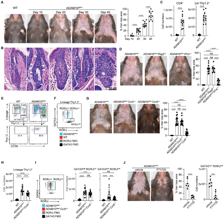
Hair follicles (HFs) function as hubs for stem cells, immune cells, and commensal microbes, which must be tightly regulated during homeostasis and transient inflammation. Here we found that transmembrane endopeptidase ADAM10 expression in upper HFs was crucial for regulating the skin microbiota and protecting HFs and their stem cell niche from inflammatory destruction. Ablation of the ADAM10-Notch signaling axis impaired the innate epithelial barrier and enabled Corynebacterium species to predominate the microbiome. Dysbiosis triggered group 2 innate lymphoid cell-mediated inflammation in an interleukin-7 (IL-7) receptor-, S1P receptor 1-, and CCR6-dependent manner, leading to pyroptotic cell death of HFs and irreversible alopecia. Double-stranded RNA-induced ablation models indicated that the ADAM10-Notch signaling axis bolsters epithelial innate immunity by promoting β-defensin-6 expression downstream of type I interferon responses. Thus, ADAM10-Notch signaling axis-mediated regulation of host-microbial symbiosis crucially protects HFs from inflammatory destruction, which has implications for strategies to sustain tissue integrity during chronic inflammation.

Keywords: ADAM10; Notch; alopecia; caspase; cicatricial alopecia; dysbiosis; hair follicles; innate lymphoid cells; pyroptosis; skin microbiota.


Conflict of interest statement

Declaration of interests The authors declare no competing interests.

Figures

Fig. 1.
Fig. 1.. Ablation of Adam10 triggers an inflammatory transcriptome.
(A) Representative immunofluorescence microscopy of HF subsets in C57BL/6 mice. (B) Representative flow cytometry and frequencies of bulge, isthmus (ISM), and infundibulum (IFD). (C) Flow cytometry and quantification of YFP expression by HF subsets in ROSA26YFPΔMx1 mice (n=6, pooled from two independent experiments). (D) Adam10 read counts from bulk RNA-seq analysis of sorted uHFs YFP+ cells from induced ROSA26YFPΔMx1 and ADAM10;ROSA26YFPΔMx1 mice (n=4 per group). Heatmaps from RNA-seq analysis for selected DEGs with annotation of associated pathways in (E and F) uHFs and (G and H) in bulge cells. (C–F) Mice were harvested 4 days post-induction.
Fig. 2.
Fig. 2.. Inflammatory ILC2s mediates HF destruction.
(A) Hair loss progression in ADAM10ΔMx1 mice (n=12) and (B) Representative H&E staining of skin sections at indicated days post-induction. Arrowheads depict lymphocytic infiltrates. (C) Quantification of indicated cell types by flow cytometry (n=12 per group). (D) Gross phenotype and hair loss area in indicated mice (n=8–12 per group). (E) Flow cytometry for CCR6 on gated lineage Thy1+ ILC populations. (F) Representative flow cytometry plot of GATA3highRORγtlow-mid ILC2s and GATA3lowRORγthigh ILC3s in indicated mice. FMO: fluorescence minus one. (G) Gross phenotype and hair loss area in indicated mice (n=17 per group). (H) Quantification of lineageThy1+ ILC populations in indicated mice. (I) Flow cytometry and quantification of GATA3high RORγtmid and GATA3highRORγtlow ILC2 subsets in indicated mice. (n=16–22 per group). (J) Gross phenotype, hair loss area, and GATA3highRORgtmid ILC2 quantification in indicated mice (n=6 per group, pooled from two independent experiments). See also Figure S1 and S2.
Fig. 3.
Fig. 3.. ADAM10ΔMx1 mice recapitulate cicatricial alopecia
(A) Representative Masson’s trichrome staining of skin sections from indicated mice. (B) Representative immunofluorescence microscopy of the HFs. CD200+CD34+ area represents the bulge. (C) Quantification of bulge cell numbers from indicated mice via flow cytometry at indicated time points (n=6–9 per group). (D) Quantification of c-kit+ melanocytes and c-kit+ CD34+ precursor numbers (n=9–10 per group, pooled from two independent experiments). (E) Unsupervised clustering analysis and t-SNE plot (left) from scRNA-seq on CD45 cells sorted from the skin of WT and ADAM10ΔMx1 mice (pooled from 3 mice in each group), color-coded based on sample origin (right). (F) Pathway analysis of DEGs between uHF clusters 2 and 4, and (G) overview heatmap annotated for selected genes. (H) Immunofluorescence microscopy for CD74 in HFs (CD45) and (I) Ki-67. (J) Representative flow cytometry for MHC ll and (K) CD200 in HFs at the indicated time points. (C and D).
Fig. 4.
Fig. 4.. Dysbiosis triggers inflammatory destruction of HFs
(A) Representative dissecting microscopic images of indicated mouse skin surfaces (upper). Dotted circles depict concretions. Gram stain in uHFs (lower). (B) Representative crystal violet and (C) propidium iodide staining of plucked hairs from indicated mice. Arrowheads depict bacteria-like elements. n=3, two independent experiments. (D) Representative skin swab and hair bacterial cultures. Colony count from skin swab cultures (n=9–15). (E) Relative abundance of major taxa via 16S rRNA sequencing analysis of skin swab samples from WT and ADAM10ΔMx1 mice with or without antibiotics. Each bar depicts one mouse, n=3-4 from two independent experiments. (F) Gross phenotype, hair loss area (n=20 per group), and total or C. mastitidis colony count from skin swab cultures of indicated mice (n=5–7 per group). (G) Quantification of relevant immune cell subsets via flow cytometry with representative plots for ILC2s in indicated mice. (H) H&E and immunofluorescence microscopy for bulge markers. Rectangle depicts the bulge. Quantification of bulge cells via flow cytometry (n=15 per group). (I) Quantification of melanocytes and its precursors from indicated mice (n=12 per group). (J) Gross phenotype, hair loss area and C. mastitidis colony count from skin swab cultures of indicated mice (n=5 per group, representative of two independent experiments). See also Figure S3 and S4.
Fig. 5.
Fig. 5.. Dysbiosis and inflammation underlie the immunological activation of HFs
(A) Unsupervised clustering analysis and t-SNE plot from scRNA-seq on CD45 cells sorted from the skin of WT and ADAM10ΔMx1 mice (antibiotic-treated and -untreated, three pooled mice from each group). (B) H2-Ab1 expression depicting inflammatory and non-inflammatory uHF clusters. Origin-based t-SNE plots and pie charts showing HF cluster frequencies. Overview heatmap (C) and (D) those for selected genes in uHFs from indicated mice. (E) Flow cytometry for MHC II+ uHF cells from indicated mice and quantification (n=20 per group). (F) Quantification of MHC II+ uHF cells via flow cytometry from indicated mice (n=8–20 per group).
Fig. 6.
Fig. 6.. ADAM10-Notch signaling axis regulates host-microbial symbiosis
(A) Gross phenotype and hair loss area in indicated mice (n=9–12). (B) Total colony counts from skin (n=9–15) and (C) 16S microbiome analysis on skin swabs from indicated mice (n=6–7). (D) Gross phenotypes and hair loss area (n=11) and (E) quantification of MHC II+ uHF and bulge cells from indicated mice (n=10–15). (F) scRNA-seq analysis of uHFs. Venn diagrams show the number of overlapping genes from indicated comparisons. (G) Heatmap of core 286 genes from (F) in indicated mice, annotated for selected genes and (H) pathway analysis. (I) Venn diagram showing overlapping genes from indicated comparisons and the list of dysregulated genes. Type I IFN induced genes were identified by bulk RNAseq from infundibulum of WT mice with or without poly(I:C) injection (n=4). (J) Bactericidal activity of mBD-6 and 1, hBD-1 and −3 against C. mastitidis (n=3, two independent experiments). Defb6 mRNA expression after siRNA knockdown of indicated genes in (K) sorted uHF keratinocytes and (L, M) MPEK-BL6 cells (n=3, two independent experiments). (N) Gross phenotype, hair loss area and C. mastitidis colony count from skin swab cultures of ADAM10ΔMx1 mice treated with topical mBD-6 or vehicle (n=5, pooled from two independent experiments).
Fig. 7.
Fig. 7.. Inflammatory caspases execute HF destruction
(A) Representative immunofluorescence microscopy and immunohistochemistry for indicated molecules. Casp: caspase; Gsdm: gasdermin (B) Western blot analysis for indicated molecules from isolated WT and ADAM10ΔMx1 mouse keratinocytes (n=2, three mouse skin samples were for each group. Representative of two independent experiments) . (C) Morphology of MPEK-BL6 cells treated with ILC-derived cytokine cocktails, HKCM, or both. Arrowheads depict pyroptotic cells. Representative of >5 experiments. (D) Real-time PCR for Casp4 in MPEK-BL6 cells treated with cytokine cocktail, HCKM, or both and (E) IL-1α, IL-1β, and Il-18 quantification via ELISA in culture supernatant (n=3, two independent experiments). (F) IL-1α quantification in culture supernatant of MPEK-BL6 cells treated with HKCM and caspase-4 inhibitor, Z-YVAD-FMK (n=3, two independent experiments). (G) Gross phenotype and hair loss area of indicated mice (n=5–8 per group, pooled from two independent experiments).

Comment in

References

    1. Abu Khweek A, and Amer AO (2020). Pyroptotic and non-pyroptotic effector functions of caspase-11. Immunol. Rev 297, e12910. - PMC - PubMed
    1. Adachi T, Kobayashi T, Sugihara E, Yamada T, Ikuta K, Pittaluga S, Saya H, Amagai M, and Nagao K (2015). Hair follicle-derived IL-7 and IL-15 mediate skin-resident memory T cell homeostasis and lymphoma. Nat. Med 21, 1272–1279. - PMC - PubMed
    1. Adler BL, and Christopher-Stine L (2018). Triggers of inflammatory myopathy: insights into pathogenesis. Discov Med 25, 75–83. - PMC - PubMed
    1. Ali RS, Falconer A, Ikram M, Bissett CE, Cerio R, and Quinn AG (2001). Expression of the peptide antibiotics human beta defensin-1 and human beta defensin-2 in normal human skin. J. Invest. Dermatol 117, 106–111. - PubMed
    1. Almazán-Fernández FM, and Fernández-Crehuet Serrano P (2017). Trichomycosis axillaris dermoscopy. Dermatol. Online J 23. - PubMed

MeSH terms

Supplementary concepts