. 2022 Jan:23:184-194.
doi: 10.1016/j.jtos.2021.09.007. Epub 2021 Sep 25.

SARS-CoV-2 and its beta variant of concern infect human conjunctival epithelial cells and induce differential antiviral innate immune response

Affiliations

SARS-CoV-2 and its beta variant of concern infect human conjunctival epithelial cells and induce differential antiviral innate immune response

Sneha Singh et al. Ocul Surf. 2022 Jan.

Abstract

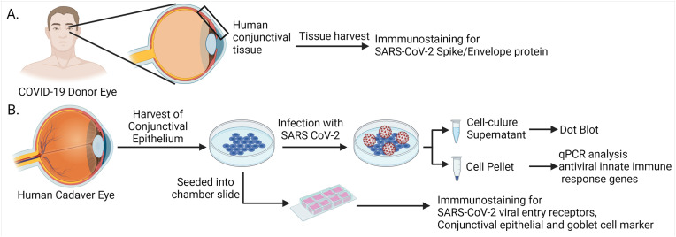
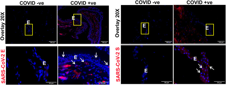
Purpose: SARS-CoV-2 RNA has been detected in ocular tissues, but their susceptibility to SARS-CoV-2 infection is unclear. Here, we tested whether SARS-CoV-2 can infect human conjunctival epithelial cells (hCECs) and induce innate immune response.

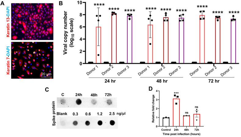
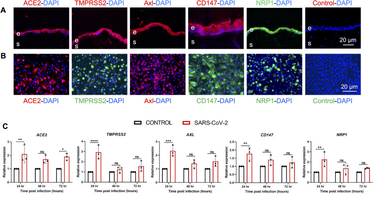
Methods: Conjunctival tissue from COVID-19 donors was used to detect SARS-CoV-2 spike and envelope proteins. Primary hCECs isolated from cadaver eyes were infected with the parental SARS-CoV-2 and its beta variant of concern (VOC). Viral genome copy number, and expression of viral entry receptors, TLRs, interferons, and innate immune response genes were determined by qPCR. Viral entry receptors were examined in hCECs and tissue sections by immunostaining. Spike protein was detected in the cell culture supernatant by dot blot.

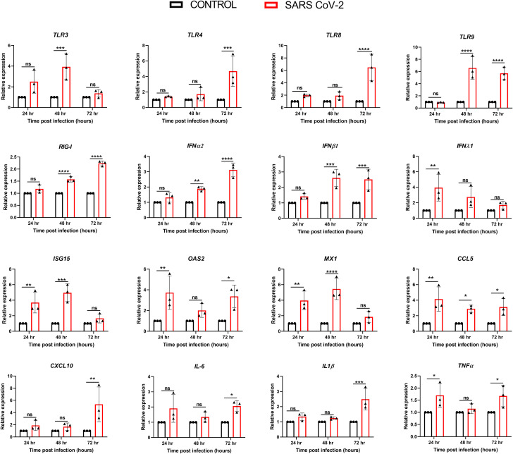
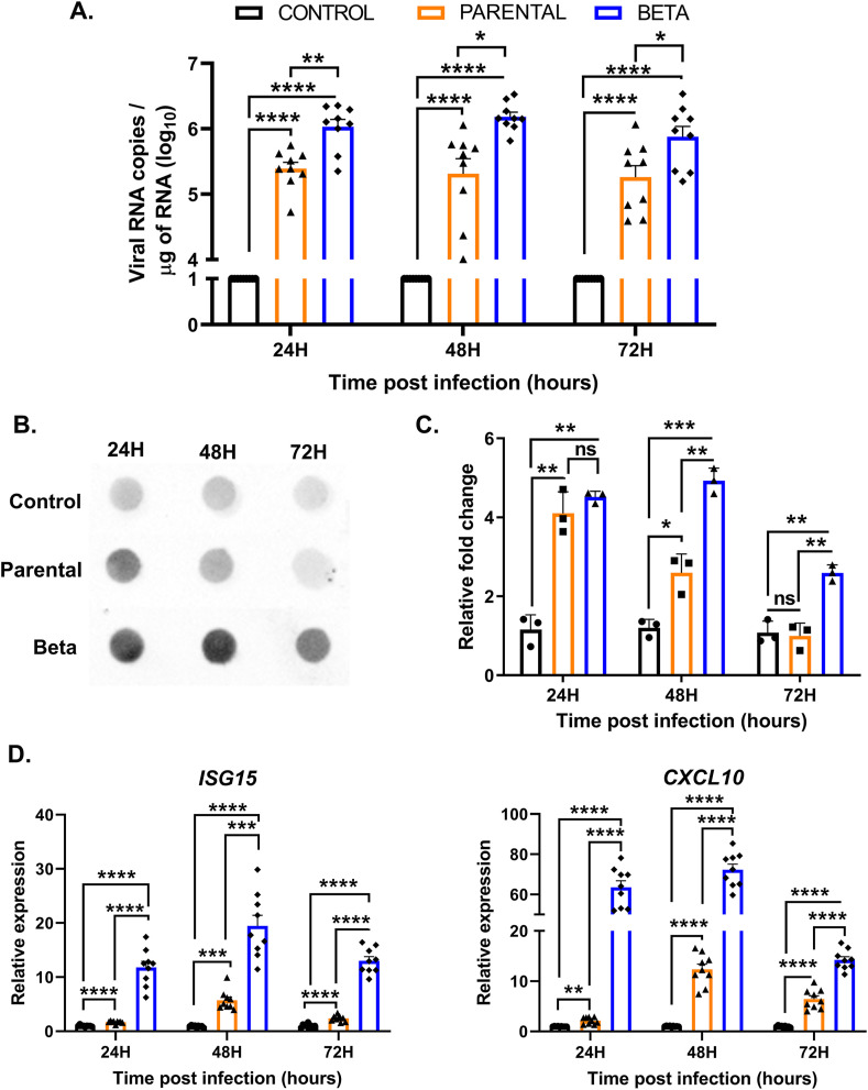
Results: Spike and envelope proteins were found in conjunctiva from COVID-19 patients. SARS-CoV-2 infected hCECs showed high viral copy numbers at 24-72h post-infection; spike protein levels were the highest at 24hpi. Viral entry receptors ACE2, TMPRSS2, CD147, Axl, and NRP1 were detected in conjunctival tissue and hCECs. SARS-CoV-2 infection-induced receptor gene expression peaked at early time points post-infection, but gene expression of most TLRs peaked at 48 or 72hpi. SARS-CoV-2 infected hCECs showed higher expression of genes regulating antiviral response, RIG-I, interferons (α, β, & λ), ISG15 & OAS2, cytokines (IL6, IL1β, TNFα), and chemokines (CXCL10, CCL5). Compared to the parental strain, beta VOC induced increased viral copy number and innate response in hCECs.

Conclusions: Conjunctival epithelial cells are susceptible to SARS-CoV-2 infection. Beta VOC is more infectious than the parental strain and evokes a higher antiviral and inflammatory response.

Keywords: COVID-19; Conjunctiva; Eye; Inflammation; Ocular surface; SARS-CoV-2; Viral entry receptors.


Conflict of interest statement

All authors declare no conflict of interest.

Figures

Fig. 1
Fig. 1
Schematic of the study design. (A) Conjunctival tissues recovered from healthy, and COVID-19 affected human donor eyes were used for immunostaining for viral proteins and viral entry receptors. (B) The primary human conjunctival epithelial cells (hCECs) were isolated from human cadaver eyes. The hCECs and ex vivo tissues were immunostained for the viral entry receptors and conjunctival epithelial and goblet cell markers. Cultured cells were infected with live SARS-CoV-2 and qPCR analysis was performed for innate immune response genes. Culture supernatant was used for dot blot assay. Images were prepared using BioRender.
Fig. 2
Fig. 2
COVID-19 donor conjunctiva showed the presence of SARS-CoV-2 envelope and spike proteins. Conjunctival tissue from healthy and COVID-19 donors was fixed in formaldehyde and 10 μm thin sections were stained for IHC using the antibody against (A) SARS-CoV-2 envelope (E) and (B) spike (S) protein (red color). DAPI was used for nuclear staining (blue color). The image was captured at different magnifications (20X and 60X) to visualize the cellular location of the viral proteins. The region of interest has been highlighted using a yellow box and white arrows. E, conjunctival epithelium.
Fig. 3
Fig. 3
Primary human conjunctival epithelial cells (hCECs) are permissive to SARS-CoV-2 infection. (A) Primary hCEC were isolated from donor eye globes and were characterized by the presence of conjunctival markers, keratins 7 and 13, by immunofluorescence staining. (B) hCECs from three donors in quadruplicate were infected with SARS-CoV-2 strain parental USA-WA1/2020 (MOI of 1) for indicated time points. qPCR analysis was performed to quantitate the viral RNA copy number wherein uninfected cells served as mock controls. (C) The cell culture supernatant from SARS-CoV-2 infected hCECs and the mock-infected hCECs was used for dot-blot assay to detect the presence of SARS-CoV-2 spike protein along with a gradient of spike protein as standard protein for quantification. The viral RNA copy number was calculated using a standard curve prepared from serial 10-fold diluted SARS-CoV-2 RNA by TaqMan based qPCR. (D) The intensity of dots was quantified using Image J software and relative fold change is plotted on the graph. One-way ANOVA; ns, non-significant; ***p < 0.001; ****p < 0.0001.
Fig. 4
Fig. 4
SARS-CoV-2 modulated the expression of viral entry receptor genes. (A) Ex vivo conjunctival tissues were immunostained for SARS-CoV-2 viral entry receptors ACE2, TMPRSS2, Axl, CD147, and NRP1. For negative control, the primary antibody was omitted and DAPI was used to stain nuclei. (B) The primary hCECs were immunostained for indicated viral entry receptors, nuclei were counterstained with DAPI. (C) hCECs isolated from three human donor cadaver eyes were infected with SARS-CoV-2 parental strain USA-WA1/2020 (MOI of 1) for indicated time points. qPCR analysis was performed to check the expression of ACE2, TMPRSS2, CD147, NRP1, and AXL genes. The experiment was performed in biological triplicates and technical duplicates and statistical analysis was performed using one-way ANOVA; ns, non-significant; *p < 0.05; **p < 0.01; ***p < 0.001; ****p < 0.0001.
Fig. 5
Fig. 5
SARS-COV-2 evoked antiviral innate and inflammatory response in hCECs. hCECs isolated from three human donor cadaver eyes were infected with SARS-CoV-2 parental strain USA-WA1/2020 (MOI of 1) for 24, 48, and 72h. qPCR analysis was performed for temporal expression of genes encoding TLRs (TLR 3, 4, 8, and 9), RIGI, IFNs (IFNα2, IFNβ1, and IFNλ1), IFN-induced antiviral response genes (ISG15, OAS2, and MX1), and inflammatory mediators (CCL5, CXCL10,IL6, IL1β, and TNFα). The experiment was performed in biological triplicates and statistical analysis was performed using one-way ANOVA; ns non-significant; *p < 0.05; **p < 0.01; ***p < 0.001; ****p < 0.0001.
Fig. 6
Fig. 6
Beta variant of SARS-COV-2 exhibited increased infectivity and higher induction of antiviral and inflammatory responses. hCECs from three donors were infected with SARS-CoV-2 parental strain and beta variant at a lower MOI of 0.1 for indicated time points. (A) qPCR analysis was performed to quantitate the viral RNA copy number wherein uninfected cells served as mock controls. (B) Culture media were used for dot blot detection of SARS-CoV-2 spike protein and the densitometry values were plotted with respect to the mock-infected control (C). (D) The expression of select antiviral response genes, ISG15, and inflammatory cytokine, CXCL10, was quantitated by qPCR. The experiment was performed in biological triplicates and statistical analysis was performed using one-way ANOVA; ns non-significant; *p < 0.05; **p < 0.01; ***p < 0.001; ****p < 0.0001.

References

    1. Guan W.J., Ni Z.Y., Hu Y., Liang W.H., Ou C.Q., He J.X., et al. Clinical Characteristics of coronavirus disease 2019 in China. N Engl J Med. 2020;382:1708–1720. - PMC - PubMed
    1. Mahase E. Covid-19: WHO declares pandemic because of "alarming levels" of spread, severity, and inaction. BMJ. 2020;368:m1036. - PubMed
    1. Ramaiah A., Arumugaswami V. Insights into cross-species evolution of novel human coronavirus SARS-CoV-2 and defining immune determinants for vaccine development. BioRxiv. 2021 doi: 10.1101/2020.01.29.925867. - DOI
    1. Medicine JHUo. CORONAVIRUS RESEARCH CENTER . John Hopkins University of Medicine: John Hopkins University of Medicine; 2021.
    1. Abdelrahman Z., Li M., Wang X. Comparative review of SARS-CoV-2, SARS-CoV, MERS-CoV, and influenza A respiratory viruses. Front Immunol. 2020;11 - PMC - PubMed

Publication types

Supplementary concepts