. 2021 Sep 28;11(1):19203.
doi: 10.1038/s41598-021-98840-w.

Enhancing esophageal repair with bioactive bilayer mesh containing FGF

Affiliations

Enhancing esophageal repair with bioactive bilayer mesh containing FGF

Ozkan Cesur et al. Sci Rep. .

Abstract

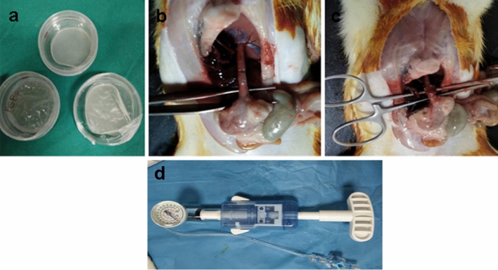
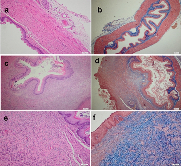
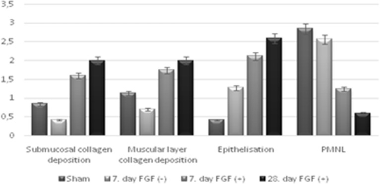
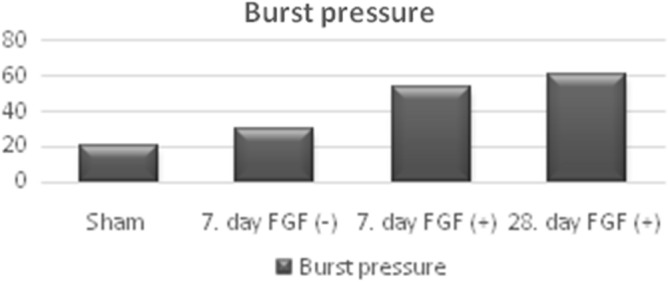
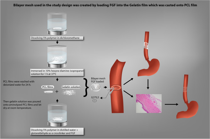
We aimed to prepare a bioactive and biodegradable bilayer mesh formed by fibroblast growth factor (FGF) loaded gelatin film layer, and poly ε-caprolactone (PCL) film layer, and to investigate its treatment efficacy on esophageal anastomosis. It is envisaged that the bioactive mesh in in vivo model would improve tissue healing in rats. The full thickness semicircular defects of 0.5 × 0.5 cm2 were created in anterior walls of abdominal esophagus. The control group had abdominal esophagus isolated with distal esophageal blunt dissection, and sham group had primary anastomosis. In the test groups, the defects were covered with bilayer polymeric meshes containing FGF (5 μg/2 cm2), or not. All rats were sacrificed for histopathology investigation after 7 or 28 days of operation. The groups are coded as FGF(-)-7th day, FGF(+)-7th day, and FGF(+)-28th day, based on their content and operation day. Highest burst pressures were obtained for FGF(+)-7th day, and FGF(+)-28th day groups (p < 0.005) and decreased inflammation grades were observed. Submucosal and muscular collagen deposition scores were markedly increased in these groups compared to sham and FGF(-)-7th day groups having no FGF (p = 0.002, p = 0.001, respectively). It was proved that FGF loaded bioactive bilayer mesh provided effective repair, reinforcement and tissue healing of esophageal defects.


Conflict of interest statement

The authors declare no conflicts of interest. And there has been no significant financial support for this work that could have influenced its outcomes.

Figures

Figure 1
Figure 1
Design of study (a) bilayer meshes, (b) preparation of esophagial defect, (c) the mesh was placed as a patch over the esophageal opening, then the edges of the esophageal opening were secured with interrupted sutures, (d) syhngomanometer device.
Figure 2
Figure 2
Mean histopathological pictogrammes of the groups studied. (a, b) Esophageal mucosa belonging to the control group. (a) There is normal archetype preserved esophageal full-thickness mucosa sample containing lamina propria and muscularis propria consisting of compact connective tissue observed under the surface ceratinized squamous epithelium, in the Hematoxylin&Eosin (H&E), × 100). (b) There is no collagen increase in the submucosal and muscular layers, in the Mason Trichrome Stain, × 100. (c, d) Esophageal mucosa belonging to the groups of sham and FGF(−). For the Groups of Sham and FGF(−) not received FGF, there are noteworthy epithelial degeneration, intense inflammation in the submucosa and muscular layer, and mildly increased collagen, in the Mason Trichrome Stain, × 100 and Hematoxylin&Eosin (H&E), × 100. (e, f) Esophageal mucosa belonging to the groups given FGF. (e) In the surface epithelium, there is marked regeneration as well as the presence of significantly increased collagen, which is replaced by the submucosal and muscular layer. There is a moderate decrease in the inflammatory cell density observed between the collagen bundles on the 7th day and a significant decrease on the 28th day, in the Hematoxylin&Eosin (H&E), × 100. (f) There is significantly increased collagen (blue color) between the submucosal and muscular layers observed, in the Mason Trichrome Stain, × 100.
Figure 3
Figure 3
Mean histopathological scores according to groups.
Figure 4
Figure 4
Differences of mean bursting pressure in the groups.
Figure 5
Figure 5
Bilayer mesh used in the study design was created by loading FGF into the Gelatin film which was casted onto PCL film showing preparation of bilayer meshes; PCL films were prepared by dissolving 5% polymer in dichloromethane. Dried PCL films were immersed in 10% hexane diamine-isopropanol solution for 1 h at 37 °C for aminolization. PCL films were washed with deionized water for 24 h. Gelatin solution was prepared by dissolving 5% polymer in distilled water and with addition of glutaraldehyde as crosslinker, and FGF. Gelatin solution was poured onto aminolyzed PCL films and let dry at room temperature. A full thickness semicircular defect of 0.5 × 0.5 cm2 was created via cutting in the anterior wall of the abdominal esophagus. The defect was repaired with primary anastomosis with the prepared bilayer mesh, using interrupted sutures.

References

    1. Kunisaki SM, Coran AG. Esophageal replacement. Semin. Pediatr. Surg. 2017;26:105–115. doi: 10.1053/j.sempedsurg.2017.02.006. - DOI - PubMed
    1. Spitz L. Esophageal atresia. J. Pediatr. Surg. 2006;41:1635–1640. doi: 10.1016/j.jpedsurg.2006.07.004. - DOI - PubMed
    1. Londono R, Badylak SF. Regenerative medicine strategies for esophageal repair. Tissue Eng. Part B Rev. 2015;21:393–410. doi: 10.1089/ten.teb.2015.0014. - DOI - PMC - PubMed
    1. Langer R, Vacanti J. Tissue engineering. Science (80-). 1993;260:920–926. doi: 10.1126/science.8493529. - DOI - PubMed
    1. Vacanti JP, Langer R. Tissue engineering: the design and fabrication of living replacement devices for surgical reconstruction and transplantation. Lancet. 1999;354:S32–S34. doi: 10.1016/S0140-6736(99)90247-7. - DOI - PubMed