. 2021 Sep;53(9):1423-1436.
doi: 10.1038/s12276-021-00676-x. Epub 2021 Sep 28.

Human cardiac stem cells rejuvenated by modulating autophagy with MHY-1685 enhance the therapeutic potential for cardiac repair

Affiliations

Human cardiac stem cells rejuvenated by modulating autophagy with MHY-1685 enhance the therapeutic potential for cardiac repair

Ji Hye Park et al. Exp Mol Med. 2021 Sep.

Erratum in

Abstract

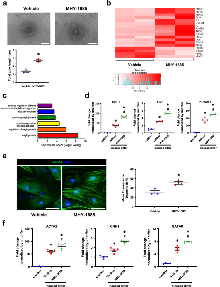
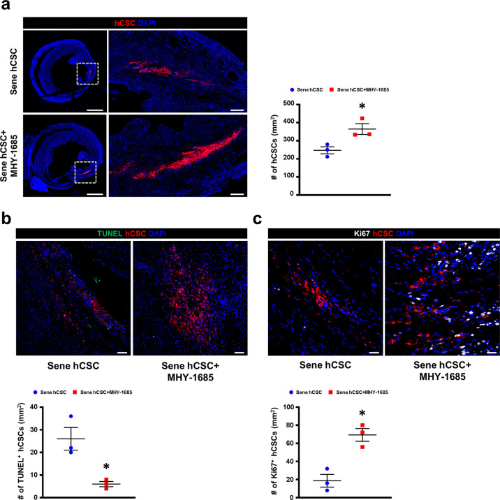
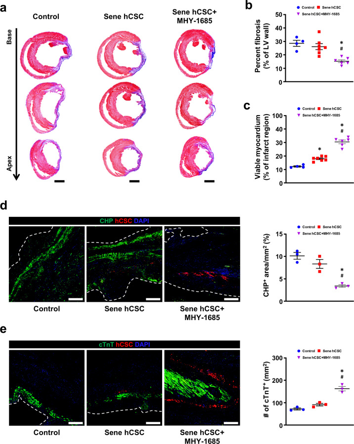
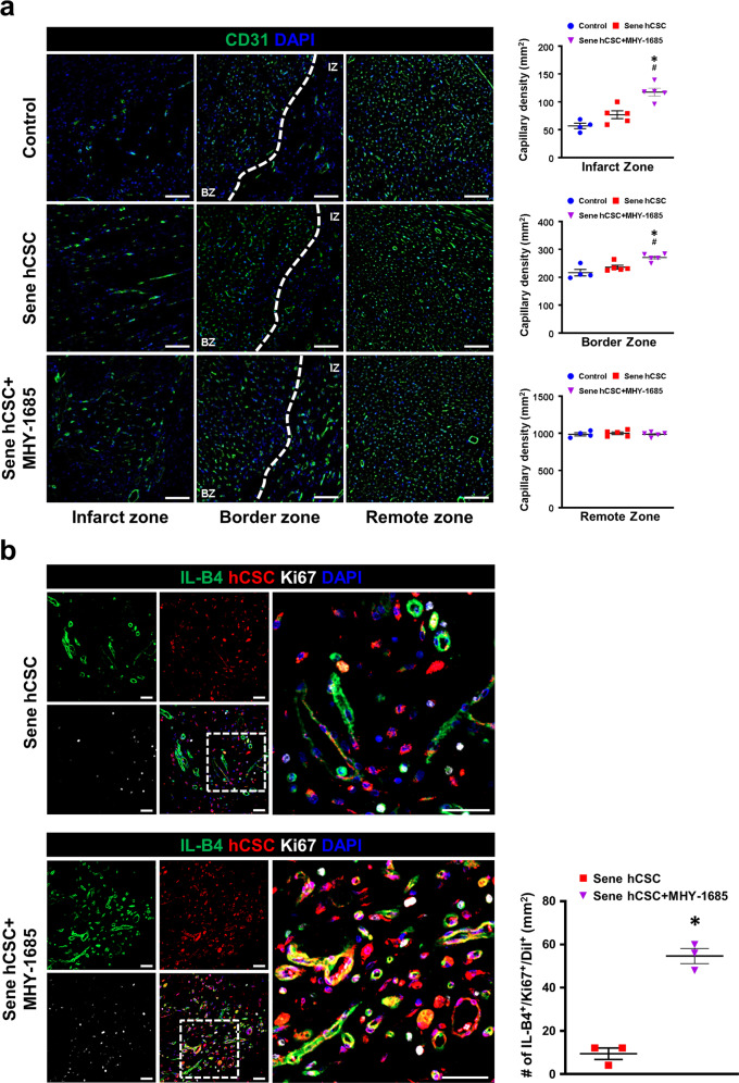
Stem cell-based therapies with clinical applications require millions of cells. Therefore, repeated subculture is essential for cellular expansion, which is often complicated by replicative senescence. Cellular senescence contributes to reduced stem cell regenerative potential as it inhibits stem cell proliferation and differentiation as well as the activation of the senescence-associated secretory phenotype (SASP). In this study, we employed MHY-1685, a novel mammalian target of rapamycin (mTOR) inhibitor, and examined its long-term priming effect on the activities of senile human cardiac stem cells (hCSCs) and the functional benefits of primed hCSCs after transplantation. In vitro experiments showed that the MHY-1685‒primed hCSCs exhibited higher viability in response to oxidative stress and an enhanced proliferation potential compared to that of the unprimed senile hCSCs. Interestingly, priming MHY-1685 enhanced the expression of stemness-related markers in senile hCSCs and provided the differentiation potential of hCSCs into vascular lineages. In vivo experiment with echocardiography showed that transplantation of MHY-1685‒primed hCSCs improved cardiac function than that of the unprimed senile hCSCs at 4 weeks post-MI. In addition, hearts transplanted with MHY-1685-primed hCSCs exhibited significantly lower cardiac fibrosis and higher capillary density than that of the unprimed senile hCSCs. In confocal fluorescence imaging, MHY-1685‒primed hCSCs survived for longer durations than that of the unprimed senile hCSCs and had a higher potential to differentiate into endothelial cells (ECs) within the infarcted hearts. These findings suggest that MHY-1685 can rejuvenate senile hCSCs by modulating autophagy and that as a senescence inhibitor, MHY-1685 can provide opportunities to improve hCSC-based myocardial regeneration.


Conflict of interest statement

The authors declare no competing interests.

Figures

Fig. 1
Fig. 1. MHY-1685 attenuated the hCSC senescent phenotype.
a Schematic diagram of the experimental design. b After a long-term culture with vehicle or MHY-1685, the relative percentage of cell viability was measured by a CCK-8 assay (*p < 0.05 vs. vehicle). c hCSCs were continuously treated with vehicle or MHY-1685, and the proliferation rate of hCSCs was measured by a CCK-8 assay (*p < 0.05 vs. vehicle). d After treatment with vehicle or MHY-1685, the S-phase cells were quantified by a BrdU incorporation assay (*p < 0.05 vs. vehicle). e Expression of the cell cycle-related proteins Cyclin E, CDK2, Cyclin D1, and CDK4 as quantified by western blot assay (*p < 0.05 vs. vehicle). f Representative images of the cell morphology of hCSCs at the same passage. The cell length and width were measured and are presented as a graph (*p < 0.05 vs. vehicle). Scale bar: 100 µm. g After long-term culture with vehicle or MHY-1685, cellular senescence was quantified by SA-β-gal-positive cells (*p < 0.05 vs. vehicle). Scale bar: 50 µm. h Population doubling time of hCSCs with vehicle or MHY-1685 (*p < 0.05 vs. vehicle). i Heat map analysis of SASP-related genes after treatment with vehicle or MHY-1685. j Expression of P16INK4a, p53, and p27 as quantified by a western blot assay (*p < 0.05 vs. vehicle). Data are shown as the mean ± S.E.M.
Fig. 2
Fig. 2. MHY-1685 improved the differentiation capacity of senescent hCSC.
a Endothelial tube formation ability of senescent hCSCs was determined by a tube formation assay (*p < 0.05 vs. vehicle). Scale bar: 50 µm. b Heatmap analysis of the positive regulation of gene expression related to angiogenesis. c Enrichment of angiogenesis-related GO terms of genes that were significantly regulated in the MHY-1685-primed hCSCs. d Expression of CDH5, Flk1, and PECAM1 after differentiation into endothelial cell lineage as examined by qRT-PCR (*p < 0.05 vs. undifferentiated hCSCs, #p < 0.05 vs. vehicle). e Vascular smooth muscle cell differentiation ability as evaluated by fluorescent immunocytochemistry using the myocyte marker α-SMA. α-SMA (green), DAPI (blue). (*p < 0.05 vs. vehicle). Scale bar: 50 μm. f Expression of ACTA2, CNN1, and GATA6 after differentiation into the vascular smooth muscle cell lineage as examined by qRT-PCR (*p < 0.05 vs. undifferentiated hCSCs, #p < 0.05 vs. vehicle). Data are shown as the mean ± S.E.M.
Fig. 3
Fig. 3. MHY-1685 regulates mTOR and the associated autophagy in hCSCs.
a Protein expression of autophagy markers mTOR, p-mTOR, LC3-I, LC3-II as evaluated by western blotting (*p < 0.05 vs. vehicle). b Autophagy detection with various concentrations of MHY-1685 in senescent hCSCs by CYTO-ID (*p < 0.05 vs. vehicle). c hCSCs were immunostained with anti-LC3 antibody (yellow), anti-LAMP1 (red), and DAPI (blue) to identify the occurrence of autophagy after treatment with MHY-1685. Scale bar: 50 μm. d The proliferative ability of senescent hCSCs (vehicle, 1 µM MHY-1685-treated hCSCs, 1 µM MHY-1685 + 20 µM + CQ-treated hCSCs) measured by a CCK-8 assay after treatment with MHY-1685 + CQ (*p < 0.05 vs. vehicle, #p < 0.05 vs. MHY-1685) e The senescence of senescent hCSCs was verified by counting the SA-β-gal-positive cells after treatment with MHY-1685 + CQ (*p < 0.05 vs. vehicle, #p < 0.05 vs. MHY-1685). f The differential ability of senescent hCSCs into ECs was verified by a tube formation assay after treatment with MHY-1685 + CQ (*p < 0.05 vs. vehicle, #p < 0.05 vs. MHY-1685). g Representative image of the differential ability of senescent hCSCs to differentiate into SMCs after treatment with MHY-1685 + CQ. α-SMA (green), DAPI (blue). Scale bar: 100 µm. h The differential ability of senescent hCSCs into SMCs was evaluated by qRT-PCR (*p < 0.05 vs. vehicle, #p < 0.05 vs. MHY-1685). Data are shown as the mean ± S.E.M.
Fig. 4
Fig. 4. hCSC-primed MHY-1685 enhances cell survival after hCSC transplantation.
a Representative images of hCSC retention at 1 week after hCSC transplantation and their quantification summary. hCSCs (white), DAPI (blue). n = 3. (*p < 0.05 vs. senescent hCSC). Scale bar: 2000 µm (Left), 200 µm (Right). b Representative images of the TUNEL assay in the infarct zone 1 week after MI and their quantification summary. TUNEL (green), hCSC (red), DAPI (blue). n = 3. (*p < 0.05 vs. senescent hCSC). Scale bar: 50 µm. c Representative images of proliferative hCSCs stained with Ki-67 in the infarct zone 1 week after MI and their quantification summary. Ki-67 (white), hCSC (red), DAPI (blue). n = 3. (*p < 0.05 vs. senescent hCSC). Scale bar: 20 µm. Data are shown as the mean ± S.E.M.
Fig. 5
Fig. 5. MHY-1685-primed hCSCs improve heart function while reducing adverse remodeling.
a Representative images of the M-mode of four experimental groups at 1 and 4 weeks postintervention. b Left ventricular ejection fraction (EF), c Left fractional shortening (FS), d Left ventricular internal diastolic dimension (LVIDd), e Left ventricular internal systolic dimension (LVIDs), f Relative wall thickness (RWT), g Septal wall thickness (SWT), h Posterior wall thickness (PWT). n = 6. (*p < 0.05 vs. control, #p < 0.05 vs. senescent hCSC). Data are shown as the mean ± S.E.M.
Fig. 6
Fig. 6. hCSC-primed MHY-1685 reduces adverse cardiac remodeling and protects the myocardium against ischemic injury.
a Representative images of Masson’s trichrome staining using heart tissues harvested 4 weeks after intervention. Scale bar: 2000 µm. b, c Quantification summary of the percentage of fibrosis and viable myocardium. n = 4–7. (*p < 0.05 vs. control, #p < 0.05 vs. senescent hCSC). d Representative images of denatured collagen in the infarct zone and their quantification summary. CHP (green), DiI-labeled hCSCs (red), DAPI (blue). n = 3. (*p < 0.05 vs. control, #p < 0.05 vs. senescent hCSC). Scale bar: 100 µm. e Representative images of cardiomyocytes in the infarct zone and their quantification summary. cTnT (green), DiI-labeled hCSCs (red), DAPI (blue). n = 3. (*p < 0.05 vs. control, #p < 0.05 vs. senescent hCSC). Scale bar: 100 µm. Data are shown as the mean ± S.E.M.
Fig. 7
Fig. 7. MHY-1685-primed hCSCs improve vascular regeneration through angiogenesis and differentiate into ECs.
a Representative images of the capillary density in the infarct zone, border zone, and remote zone 4 weeks after MI and their quantification summary. CD31 (green), DAPI (blue). n = 4–5. (*p < 0.05 vs. control, #p < 0.05 vs. senescent hCSC). Scale bar: 100 µm. b Representative images of hCSCs differentiated into ECs and proliferation in the infarct zone and quantification summary. IL-B4 (green), DiI-labeled hCSCs (red), Ki-67 (white), DAPI (blue). n = 3. (*p < 0.05 vs. senescent hCSC). Scale bar: 30 µm. Data are shown as the mean ± S.E.M.

References

    1. Thygesen K, et al. Fourth universal definition of myocardial infarction (2018) J. Am. Coll. Cardiol. 2018;72:2231–2264. doi: 10.1016/j.jacc.2018.08.1038. - DOI - PubMed
    1. Dimmeler S, Burchfield J, Zeiher AM. Cell-based therapy of myocardial infarction. Arterioscler. Thromb. Vasc. Biol. 2008;28:208–216. doi: 10.1161/ATVBAHA.107.155317. - DOI - PubMed
    1. Beltrami AP, et al. Adult cardiac stem cells are multipotent and support myocardial regeneration. Cell. 2003;114:763–776. doi: 10.1016/S0092-8674(03)00687-1. - DOI - PubMed
    1. Malliaras K, et al. Safety and efficacy of allogeneic cell therapy in infarcted rats transplanted with mismatched cardiosphere-derived cells. Circulation. 2012;125:100–112. doi: 10.1161/CIRCULATIONAHA.111.042598. - DOI - PMC - PubMed
    1. Tang XL, et al. Intracoronary administration of cardiac progenitor cells alleviates left ventricular dysfunction in rats with a 30-day-old infarction. Circulation. 2010;121:293–305. doi: 10.1161/CIRCULATIONAHA.109.871905. - DOI - PMC - PubMed

Publication types

Substances