. 2021 Sep 28;4(1):1136.
doi: 10.1038/s42003-021-02653-6.

Actin crosslinker competition and sorting drive emergent GUV size-dependent actin network architecture

Affiliations

Actin crosslinker competition and sorting drive emergent GUV size-dependent actin network architecture

Yashar Bashirzadeh et al. Commun Biol. .

Abstract

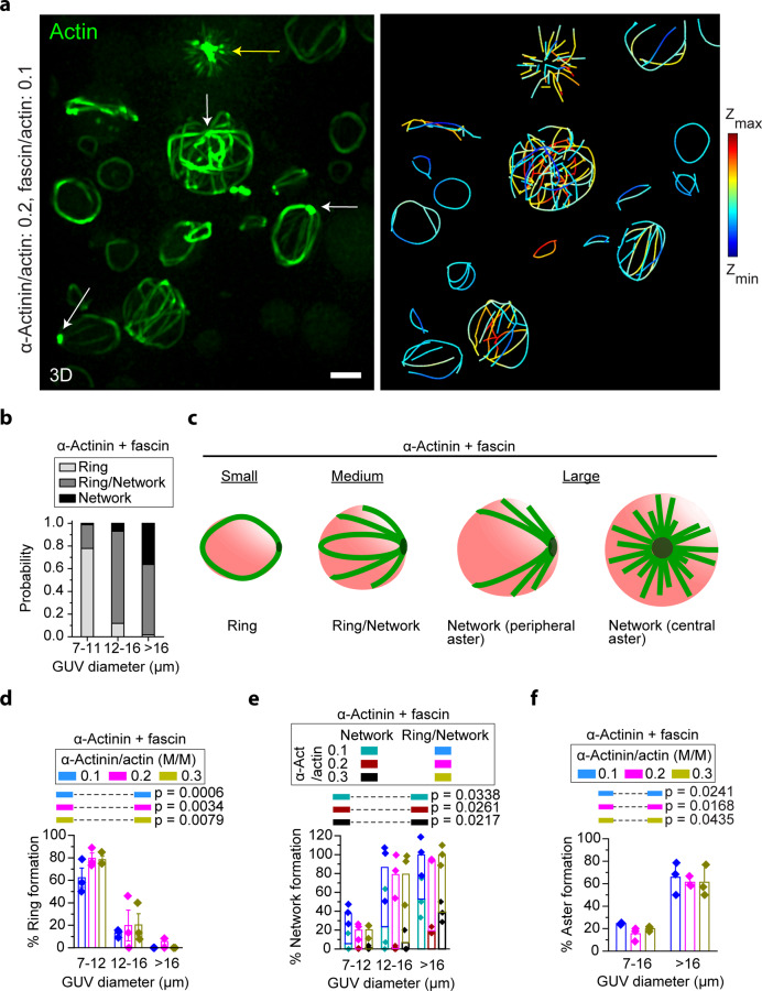
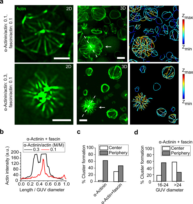
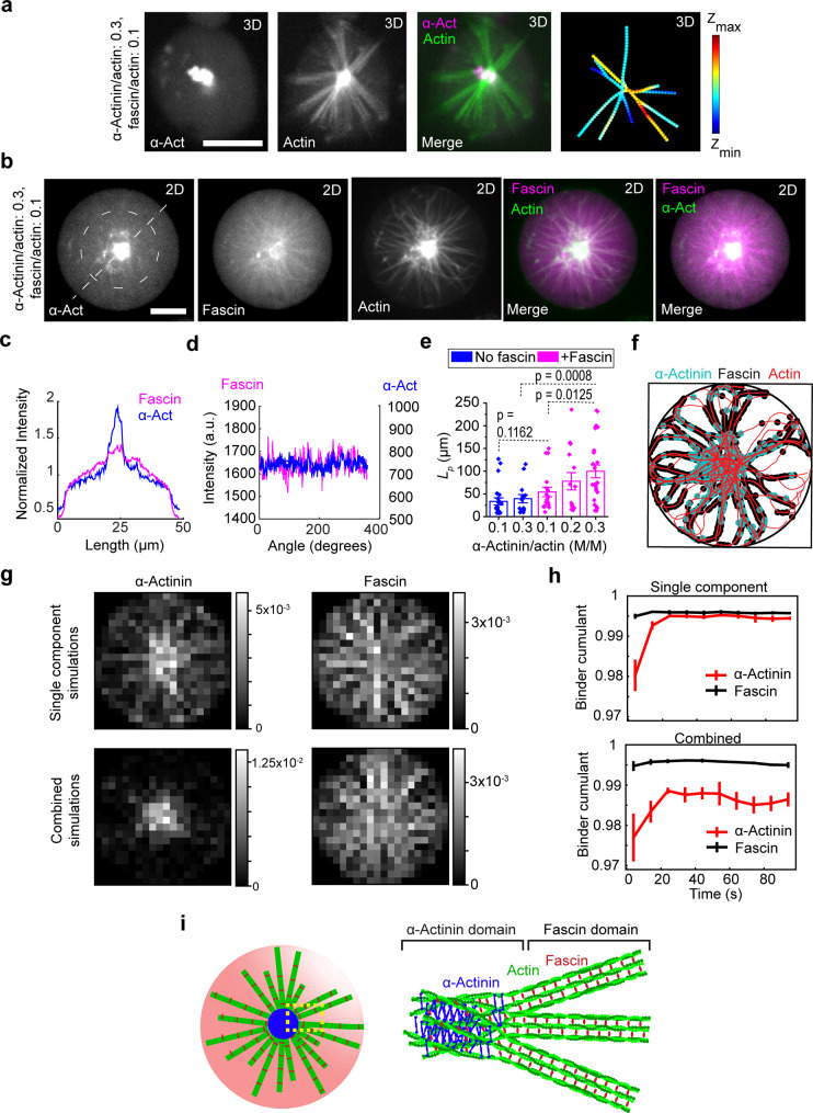
The proteins that make up the actin cytoskeleton can self-assemble into a variety of structures. In vitro experiments and coarse-grained simulations have shown that the actin crosslinking proteins α-actinin and fascin segregate into distinct domains in single actin bundles with a molecular size-dependent competition-based mechanism. Here, by encapsulating actin, α-actinin, and fascin in giant unilamellar vesicles (GUVs), we show that physical confinement can cause these proteins to form much more complex structures, including rings and asters at GUV peripheries and centers; the prevalence of different structures depends on GUV size. Strikingly, we found that α-actinin and fascin self-sort into separate domains in the aster structures with actin bundles whose apparent stiffness depends on the ratio of the relative concentrations of α-actinin and fascin. The observed boundary-imposed effect on protein sorting may be a general mechanism for creating emergent structures in biopolymer networks with multiple crosslinkers.


Conflict of interest statement

The authors declare no competing interests.

Figures

Fig. 1
Fig. 1. GUV size- and cross-linker concentration-dependent organization of actin-α-actinin networks.
a Representative 3D-reconstructed fluorescence confocal images of actin networks of different α-actinin:actin ratios (actin concentration at 5 µM). Actin bundles form networks in larger GUVs, while they form single rings in smaller GUVs. Dotted lines outline GUV boundaries. Scale bar, 10 µm. b Cumulative probability (for all three α-actinin concentrations) of ring and network formation in GUVs with different sizes. c, d Probability of the formation of rings and networks at different α-actinin concentrations. Error bars in (d) indicate standard error of the mean; n = 3 experiments. NGUVs per experiment = [389 42 23], [67 45 38], [188 145 128] in order of ascending α-actinin concentration (numbers in brackets are arranged in order of ascending GUV diameter). p values compare formation probabilities of rings (c) and networks (d) in small and large GUVs. e Representative 3D-reconstructed image from confocal stack of fluorescence images of an encapsulated α-actinin/actin (3:10 [M/M]) network. High α-actinin concentration can induce the formation of dense clusters at the GUV periphery. Scale bar, 10 µm. f The probability of the formation of actin peripheral asters depends on GUV size. The majority of actin bundles form peripheral asters in larger GUVs. Error bars indicate standard error of the mean; n = 3 experiments. NGUVs per experiment = [429 23], [112 38], [333 128] in order of ascending α-actinin concentration (numbers in brackets are arranged in order of ascending GUV diameter). p values compare peripheral aster-formation probabilities for the two ranges of GUV diameters. g Cumulative (three experiments) proportion of peripheral asters with peripheral bundles (all actin bundles elongated around GUV periphery) and luminal bundles (with at least one actin bundle elongated in GUV lumen) in large GUVs (diameter > 16 μm). At high α-actinin concentrations, the majority of actin bundles form asters with all actin bundles elongated around the periphery (peripheral bundles). Number of large GUVs with peripheral asters = [35, 14, 53] in order of ascending α-actinin concentration. h Schematic summary of the result of encapsulated actin-network assembly by α-actinin (without fascin) in different sized vesicles. Blue arrows show the merging of actin-filament bundles (green dashed lines) into distinct peripheral bundles (green solid lines).
Fig. 2
Fig. 2. GUV-size-dependent formation of rings, peripheral asters, and central asters by α-actinin and fascin.
a Representative 3D-reconstructed (left) and skeletonized (right) images from a confocal fluorescence image stack of 5 µM actin (10% ATTO-488 actin) bundled by 0.5 µM fascin and 1 µM α-actinin in GUVs (composition: 69.9% DOPC, 30% cholesterol, and 0.1% rhodamine-PE). Yellow arrow denotes a cluster of actin fluorescence in a central aster. White arrows denote clusters of actin fluorescence in peripheral asters. Color in the skeletonized image shows z position. Scale bar, 10 μm. b Cumulative probability (α-actinin concentrations of 0.5, 1, and 1.5 µM with 0.5 µM fascin and 5 µM actin) of ring and network formation in GUVs with different sizes. c Schematic representation of GUV-size-dependent actin networks assembled by α-actinin and fascin. Rings and peripheral asters can occasionally deform GUVs. d, e Probability of the formation of rings (d) and networks (e) at different α-actinin concentrations as a function of GUV diameter. NGUVs per experiment = [101 98 29], [147 58 71], [137 82 24] in order of ascending α-actinin concentration (numbers in brackets are arranged in order of ascending GUV diameter). p values compare formation probabilities of rings (d) and networks (e) in small and large GUVs. f Probability of aster formation (peripheral or central) in the presence of fascin and α-actinin. Aster formation depends on GUV size but not α-actinin concentration. Fascin/actin, 0.1 (M/M). All error bars indicate standard error of the mean; n = 3 experiments. NGUVs per experiment = [199 29], [205 71], [219 24] in order of ascending α-actinin concentration (numbers in brackets are arranged in order of ascending GUV diameter). p values compare aster-formation probabilities in the two ranges of GUV diameters.
Fig. 3
Fig. 3. GUV-size-dependent localization of F-actin clusters in the presence of α-actinin and fascin suggests cross-linker sorting and domain formation.
a Representative 2D (left) confocal fluorescence images of actin networks shown by arrows (3D-reconstructed image, middle) along with skeletonized (right) image of the GUV populations. α-actinin, 0.5 μM (top), 1.5 μM (bottom). Fascin, 0.5 μM. Actin, 5 μM. Scale bar, 10 μm. b Actin fluorescence intensity along the length of dashed lines drawn across the two GUVs in (a) normalized to the GUV diameters. c Cumulative probability (for all α-actinin concentrations) of the aggregation of cross-linked actin with/without fascin in GUVs with diameter >20 μm. α-actinin–fascin–actin bundles tend to shift cluster localization from the periphery to the center of GUVs. Probabilities do not add to 100% because a portion of actin bundles do not cluster at either the center or the periphery. NGUVs>20 μm = 167 [87 (α-actinin), 80 (α-actinin+fascin)]. In our analysis, only a minority of vesicles with encapsulated α-actinin–actin-bundle structures and encapsulated α-actinin–fascin–actin-bundle structures did not form clusters. d Cumulative probability (for all α-actinin concentrations) of the aggregation of cross-linked actin in large GUVs in the presence of both α-actinin and fascin. Larger GUVs facilitate centering of clusters. A portion of actin bundles do not cluster either at the center or at the periphery. Nanalyzed GUVs>16 μm = 87 (61 [17–24 μm], 26 [>24 μm]).
Fig. 4
Fig. 4. α-Actinin and fascin sort in central aster structures.
a Representative 3D-reconstructed confocal fluorescence images of α-actinin, actin, merged, and skeletonized construct of an encapsulated central aster, respectively, from left to right. α-Actinin, 1.5 μM (including 14 mol% TMR α-actinin). Fascin, 0.5 μM (including 50 mol% AF647 Fascin). Actin, 5 μM. Scale bar, 10 μm. b Representative 2D confocal fluorescence images of α-actinin, fascin, actin, and merged images of an encapsulated central aster in a large GUV. α-Actinin, 1.5 μM (including 13 mol% TMR α-actinin). Fascin, 0.5 μM (including 16 mol% TMR α-actinin). Actin, 5 μM. Scale bar, 10 μm. c, d Fluorescence intensity of α-actinin and fascin along the dashed line drawn across the GUV (c) and circle drawn around central aster outside the actin cluster (d) in (b). e Persistence length of actin bundles without and with fascin (fascin/actin, 0.1 [M/M]) at different α-actinin/actin ratios indicated. Lp [fascin:α-actinin:actin] = 33.3 ± 10 μm [0:1:10], 39.3 ± 9 μm [0:3:10], 54.2 ± 11.4 μm [1:1:10], 77.8 ± 19 μm [1:2:10], and 99.7 ± 15 μm [1:3:10]. Nbundles = [22 14 17 14 26] in order of x-axis categories, 3 GUVs per category. f Representative structure after 100 s of simulation of actin filaments (red) cross-linked by α-actinin (cyan) and fascin (black). The border of the containing circle is shown in black. g PDFs of α-actinin (left) and fascin (right) from either simulation of each alone with actin (top row) or the two in combination with actin (bottom row). PDFs are constructed from the last frames of five independent simulations of 100 s for each condition. h Binder cumulant as a function of time for each cross-linker separately with actin (top) and the combined simulation (bottom). Values are measured for each simulation independently and then averaged. Error bars represent one standard deviation from the mean. i Schematic of cross-linker-size-dependent sorting in confined α-actinin–fascin cross-linked actin network.

References

    1. Pegoraro AF, Janmey P, Weitz DA. Mechanical properties of the cytoskeleton and cells. Cold Spring Harb. Perspect. Biol. 2017;9:a022038. doi: 10.1101/cshperspect.a022038. - DOI - PMC - PubMed
    1. Dumont S, Prakash M. Emergent mechanics of biological structures. Mol. Biol. Cell. 2014;25:3461–3465. doi: 10.1091/mbc.e14-03-0784. - DOI - PMC - PubMed
    1. Fletcher DA, Mullins RD. Cell mechanics and the cytoskeleton. Nature. 2010;463:485–492. doi: 10.1038/nature08908. - DOI - PMC - PubMed
    1. Blanchoin L, Boujemaa-Paterski R, Sykes C, Plastino J. Actin dynamics, architecture, and mechanics in cell motility. Physiol. Rev. 2014;94:235–263. doi: 10.1152/physrev.00018.2013. - DOI - PubMed
    1. Luo T, Mohan K, Iglesias PA, Robinson DN. Molecular mechanisms of cellular mechanosensing. Nat. Mater. 2013;12:1064–1071. doi: 10.1038/nmat3772. - DOI - PMC - PubMed

Publication types

MeSH terms