Review
. 2021 Dec;21(12):786-802.
doi: 10.1038/s41568-021-00397-3. Epub 2021 Sep 28.

Glial and myeloid heterogeneity in the brain tumour microenvironment

Affiliations
Review

Glial and myeloid heterogeneity in the brain tumour microenvironment

Brian M Andersen et al. Nat Rev Cancer. 2021 Dec.

Abstract

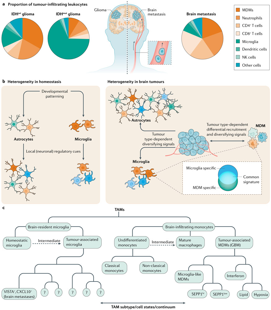
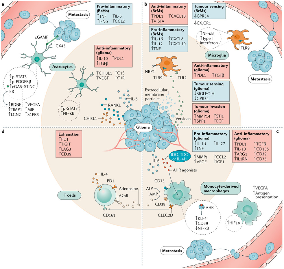
Brain cancers carry bleak prognoses, with therapeutic advances helping only a minority of patients over the past decade. The brain tumour microenvironment (TME) is highly immunosuppressive and differs from that of other malignancies as a result of the glial, neural and immune cell populations that constitute it. Until recently, the study of the brain TME was limited by the lack of methods to de-convolute this complex system at the single-cell level. However, novel technical approaches have begun to reveal the immunosuppressive and tumour-promoting properties of distinct glial and myeloid cell populations in the TME, identifying new therapeutic opportunities. Here, we discuss the immune modulatory functions of microglia, monocyte-derived macrophages and astrocytes in brain metastases and glioma, highlighting their disease-associated heterogeneity and drawing from the insights gained by studying these malignancies and other neurological disorders. Lastly, we consider potential approaches for the therapeutic modulation of the brain TME.


Figures

Fig. 1 ∣
Fig. 1 ∣. Microglia, MDM and astrocyte diversity in brain tumours.
a ∣ Pie charts represent the composition of leukocytes in isocitrate dehydrogenase wild-type (IDHwt) and IDH mutant (IDHmut) glioma and brain metastases as determined by flow cytometry and cytometry by time of flight (CyTOF). Leukocyte composition varies by tumour type,. The pie charts represent general trends from multiple tumours. b ∣ Non-malignant cell types in the tumour microenvironment (TME) have the potential for diverse activation states during both homeostasis (left panel) and cancer (right panel). Environmental cues and differential recruitment to the TME are the major drivers of heterogeneity in tumour-associated macrophages (TAMs; comprised of monocyte-derived macrophages (MDMs) and microglia). The plasticity of TME-infiltrating MDMs results in their acquisition of microglia-like transcriptional programmes. Astrocytes are highly heterogeneous, which is amplified upon activation. c ∣ Organizational structure for TAM nomenclature. Notable subsets are listed from recent reports,. Intermediate states represent subsets that exist on a continuum between undifferentiated monocytes and macrophages or homeostatic microglia and tumour-associated microglia. GBM, glioblastoma; NK, natural killer.
Fig. 2 ∣
Fig. 2 ∣. Mechanisms of cell communication involved in tumour establishment, invasion and immune escape.
Multiple signalling pathways are activated in brain tumour microenvironment astrocytes and tumour-associated macrophages, but their contribution to immunosuppression as well as the dominance of a specific pathway within specific subpopulations (not shown) are unknown. Astrocytes (part a) upregulate genes encoding immunosuppressive (IL-10, transforming growth factor-β (TGFβ) and PDL1) and pro-oncogenic (CHI3L1) factors in the glioma tumour microenvironment, while nuclear factor-κB (NF-κB) signalling is concomitantly induced by tumour-derived RANKL. STAT3 is a central regulator of astrocyte reactivity programmes and characterizes a brain metastasis-promoting astrocyte subset. Other subsets promote tumour growth through paracrine factors such as brain-derived neurotrophic factor (BDNF) or TNF through cell contact-dependent (CX43) or cell contact-independent mechanisms. Microglia (part b) and monocyte-derived macrophages (MDMs) (part c) can have both pro-inflammatory and anti-inflammatory functions. In microglia (part b), tumour sensing (SIGLEC-H and GPR34) and homeostatic (CX3C-chemokine receptor 1 (CX3CR1)) programmes are disrupted, while TLR2 and NRP1 signalling respectively increase tumour invasion and immunosuppression. In monocyte-derived macrophages (part c), an anti-inflammatory phenotype supported by aryl hydrocarbon receptor (AHR) activation suppresses T cell responses (part d) via the catabolism of ATP. T cells supply tumour-associated macrophages with a CSF1R-independent survival signal by inducing IGF1 via IL-4. Anti-tumour T cell responses are suppressed by cytokines such as IL-10 and TGFβ, checkpoint proteins such as PDL1 and VISTA, and adenosine signalling. Overall T cell exhaustion is evidenced by increased immune-checkpoint expression and the inhibitory receptors A2aR and CD161 are broadly present in glioblastoma-infiltrating T cells. ARGl, arginase 1; cGAMP, cyclic GMP–AMP; EGF, epidermal growth factor; ER, oestrogen receptor; HIF1α, hypoxia-inducible factor 1α; IGF1, insulin-like growth factor 1; MIF, macrophage inhibitory factor; MMPs, matrix metalloproteinases; NRP1, neuropilin 1; p-PDGFRβ, phosphorylated platelet-derived growth factor receptor-β; p-STAT3, phosphorylated signal transducer and activator of transcription 3; TLR, Toll-like receptor; VEGFA, vascular endothelial growth factor A.

References

    1. Tawbi HA et al. Combined nivolumab and ipilimumab in melanoma metastatic to the brain. N. Engl. J. Med 379, 722–730 (2018). - PMC - PubMed
    1. Long GV et al. Combination nivolumab and ipilimumab or nivolumab alone in melanoma brain metastases: a multicentre randomised phase 2 study. Lancet Oncol. 19, 672–681 (2018). - PubMed
    1. Goldberg SB et al. Pembrolizumab for patients with melanoma or non-small-cell lung cancer and untreated brain metastases: early analysis of a non-randomised, open-label, phase 2 trial. Lancet Oncol. 17, 976–983 (2016). - PMC - PubMed
    1. Fecci PE et al. Increased regulatory T-cell fraction amidst a diminished CD4 compartment explains cellular immune defects in patients with malignant glioma. Cancer Res. 66, 3294–3302 (2006). - PubMed
    1. Kwok D & Okada H T-cell based therapies for overcoming neuroanatomical and immunosuppressive challenges within the glioma microenvironment. J. Neurooncol 147, 281–295 (2020). - PMC - PubMed

Publication types