Review
. 2021 Sep 21:15:3877-3887.
doi: 10.2147/OPTH.S272345. eCollection 2021.

Clinical Manifestations of Cuticular Drusen: Current Perspectives

Affiliations
Review

Clinical Manifestations of Cuticular Drusen: Current Perspectives

Serena Fragiotta et al. Clin Ophthalmol. .

Abstract

Cuticular drusen are part of the spectrum of age-related macular degeneration (AMD) with particular clinical and multimodal imaging characteristics. This drusen subpopulation shares several high-risk single nucleotide polymorphisms with AMD. Despite this feature, they can manifest at a relatively young age, presenting with a female preponderance. Multimodal imaging is essential for characterizing such lesions, using a combination of color fundus photographs, optical coherence tomography (OCT), fluorescein angiography (FA), and fundus autofluorescence (FAF). The classic starry-sky pattern visible on FA and the typical central hypoautofluorescent lesion with hyperautofluorescent rim on FAF is considered the result of a central retinal pigment epithelium (RPE) erosion from these triangular elevations of the RPE-basal lamina. This finding may also be responsible for the typical choroidal hypertransmission appreciated through OCT. The clinical course of cuticular drusen may be relatively benign at early stages, with small drusen presenting at a young age. However, the presence of clinical phenotypes characterized by diffuse involvement and/or accompanying large drusen in patients older than 60 years may confer a significant risk for either macular neovascularization or geographic atrophy.

Keywords: cuticular drusen; fluorescein angiography; fundus autofluorescence; multimodal imaging; spectral-domain optical coherence tomography.


Conflict of interest statement

The authors report no conflicts of interest in this work.

Figures

Figure 1
Figure 1
Short wavelength fundus autofluorescence (FAF). (A) A 55-degree FAF acquired using a Spectralis (Heidelberg Engineering, Heidelberg, Germany) device that uses a 488 nm illumination wavelength and emission detection between 500 and 700 nm; FAF imaging demonstrated pinpoint hypoautofluorescent alterations confined within the posterior pole. (B) A 35-degree FAF magnification shows more easily the typical pattern of cuticular drusen constituted by central hypofluorescence surrounded by a hyperautofluorescent halo (yellow arrowheads, inset). (C) Subfoveal optical coherence tomography B-scan demonstrating multiple retinal pigment epithelium-basal lamina elevations with a classical saw tooth configuration.
Figure 2
Figure 2
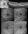
Fluorescein angiography (FA) and indocyanine green angiography (ICGA). Arteriovenous phase (0:43 seconds) showing the typical “stars-in-the-sky” appearance at both FA (A) and ICGA (B); The hyperfluorescent lesions tend to progressively fade, but persist through late phase angiograms (5:02 min) on both FA (C) and ICGA (D). (E) Infrared reflectance with scan reference (green line) of the subfoveal optical coherence tomography B-scan (F) showing multiple elevations of the retinal pigment epithelium-basal lamina with variable content.
Figure 3
Figure 3
Cuticular drusen recognition using multimodal imaging. (A) Fundus autofluorescence demonstrates multifocal central hypoautofluorescence with hyperautofluorescent border. (B) Spectral-domain optical coherence tomography (OCT) B-scan exhibiting the classical saw-tooth configuration with a hyporeflective internal content and a characteristic bar-code signature (peach arrows) in the choroid. (C) Structural en face obtained through a customized segmentation (D) passing above the plane of drusen (−95, −55 µm offsets) demonstrates their distribution. A structural en face customized view (−34, 6 µm offsets) passing through the drusen (E and F) highlights the hyporeflective cores of the cuticular drusen.
Figure 4
Figure 4
Cuticular drusen associated with large drusen. (A) Fundus autofluorescence shows the presence of large drusen appearing as faint hyperautofluorescent lesions. These large drusen are barely visible on fluorescein angiography during early phases (B) with minimal increase of hyperfluorescence during late phases (C). On spectral-domain optical coherence tomography B-scan through the foveal center, large drusen tend to coalesce into a drusenoid pigment epithelial detachment (D).
Figure 5
Figure 5
Cuticular drusen associated with vitelliform detachment. (A) Fundus autofluorescence demonstrating hyperautofluorescent signal in correspondence of vitelliform material.; (B) Subfoveal optical coherence tomography B-scan shows discrete retinal pigment epithelium-basal lamina (BL) elevations compatible with cuticular drusen and a subretinal accumulation of hyperreflective material consistent with a vitelliform detachment. (C) Fluorescein angiography (FA) at 0:50 seconds demonstrates a starry-sky appearance; this aspect is also detectable through indocyanine green angiography (ICGA) visible at (D) The starry-sky appearance is maintained in late angiograms (6:42 min) of FA (E) and ICGA (F). Additionally, late staining of vitelliform material can be appreciated with FA, while a central hypofluorescence is visible with ICGA.

References

    1. Gass JDM. Stereoscopic Atlas of Macular Diseases. Diagnosis and Treatment. Mosby CV, 2nd ed. St.Louis; 1977
    1. Gass JD, Jallow S, Davis B. Adult vitelliform macular detachment occurring in patients with basal laminar drusen. Am J Ophthalmol. 1985;99(4):445–459. doi:10.1016/0002-9394(85)90012-1 - DOI - PubMed
    1. Russell SR, Mullins RF, Schneider BL, Hageman GS. Location, substructure, and composition of basal laminar drusen compared with drusen associated with aging and age-related macular degeneration. Am J Ophthalmol. 2000;129(2):205–214. doi:10.1016/S0002-9394(99)00345-1 - DOI - PubMed
    1. Sarks SH, Arnold JJ, Killingsworth MC, Sarks JP. Early drusen formation in the normal and aging eye and their relation to age related maculopathy: a clinicopathological study. Br J Ophthalmol. 1999;83(3):358–368. doi:10.1136/bjo.83.3.358 - DOI - PMC - PubMed
    1. Farkas TG, Krill AE, Sylvester VM, Archer D. Familial and secondary drusen: histologic and functional correlations. Trans Am Acad Ophthalmol Otolaryngol. 1971;75(2):333–343. - PubMed

LinkOut - more resources