

MRI studies of brain size and growth in individuals with congenital heart disease
- PMID: 34584889
- PMCID: PMC8429874
- DOI: 10.21037/tp-20-282
MRI studies of brain size and growth in individuals with congenital heart disease
Abstract
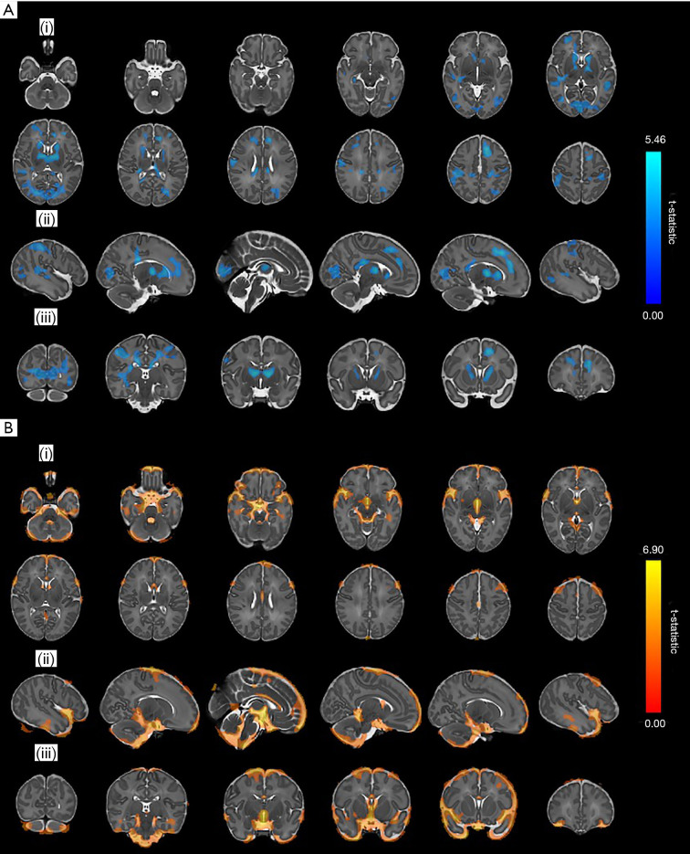
Congenital heart disease (CHD) is the most frequent congenital abnormality. Most infants born with CHD now survive. However, survivors of CHD are at increased risk of neurodevelopmental impairment, which may be due to impaired brain development in the fetal and neonatal period. Magnetic resonance imaging (MRI) provides objective measures of brain volume and growth. Here, we review MRI studies assessing brain volume and growth in individuals with CHD from the fetus to adolescence. Smaller brain volumes compared to healthy controls are evident from around 30 weeks gestation in fetuses with CHD and are accompanied by increased extracerebral cerebrospinal fluid. This impaired brain growth persists after birth and throughout childhood to adolescence. Risk factors for impaired brain growth include reduced cerebral oxygen delivery in utero, longer time to surgery and increased hospital stay. There is increasing evidence that smaller total and regional brain volumes in this group are associated with adverse neurodevelopmental outcome. However, to date, few studies have assessed the association between early measures of cerebral volume and neurodevelopmental outcome in later childhood. Large prospective multicentre studies are required to better characterise the relationship between brain volume and growth, clinical risk factors and subsequent cognitive, motor, and behavioural impairments in this at-risk population.
Keywords: Congenital heart disease (CHD); brain; brain volume; magnetic resonance imaging (MRI).
2021 Translational Pediatrics. All rights reserved.
Conflict of interest statement
Conflicts of Interest: The authors have completed the ICMJE uniform disclosure form (available at http://dx.doi.org/10.21037/tp-20-282). The series “Pre-natal Diagnosis in Congenital Heart Defects” was commissioned by the editorial office without any funding or sponsorship. IHXN reports other from National Institute for Health Research, outside the submitted work. SJC reports grants from Medical Research Council (UK), grants from Action for Medical Research, grants from British Heart Foundation, during the conduct of the study. The authors have no other conflicts of interest to declare.
Figures
References
Publication types
Grants and funding
LinkOut - more resources
Full Text Sources