Review
. 2021 Aug;10(8):2182-2196.
doi: 10.21037/tp-20-224.

Fetal brain issues in congenital heart disease

Affiliations
Review

Fetal brain issues in congenital heart disease

Fu-Tsuen Lee et al. Transl Pediatr. 2021 Aug.

Abstract

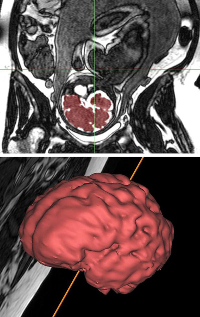
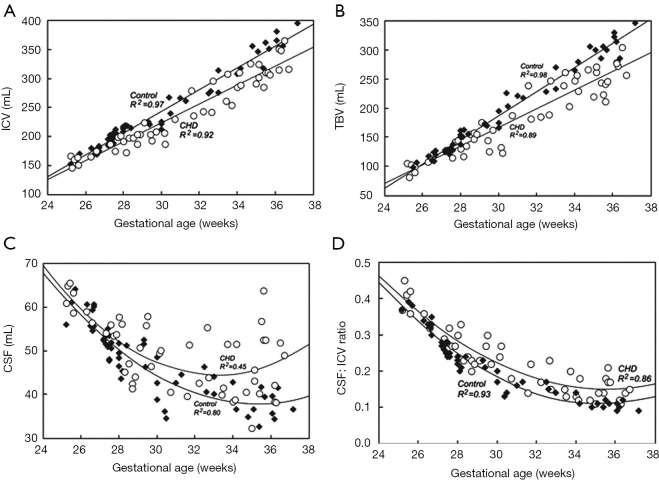
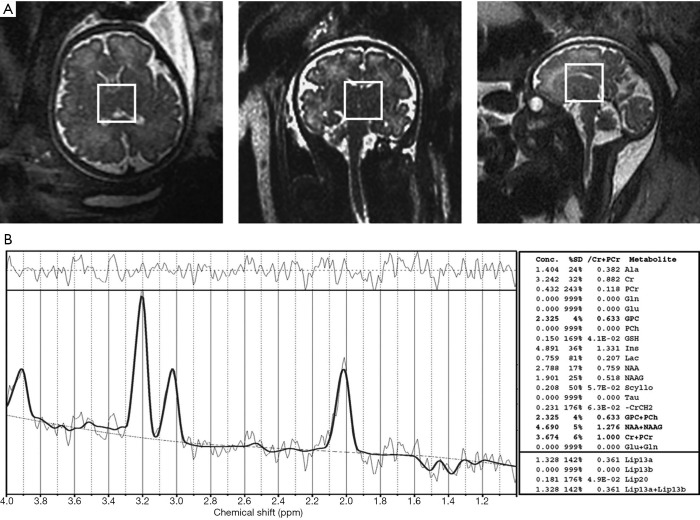
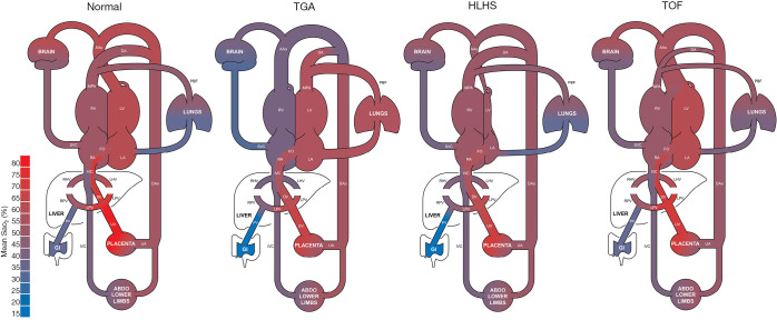
Following the improvements in the clinical management of patients with congenital heart disease (CHD) and their increased survival, neurodevelopmental outcome has become an emerging priority in pediatric cardiology. Large-scale efforts have been made to protect the brain during the postnatal, surgical, and postoperative period; however, the presence of brain immaturity and injury at birth suggests in utero and peripartum disturbances. Over the past decade, there has been considerable interest and investigations on fetal brain growth in the setting of CHD. Advancements in fetal brain imaging have identified abnormal brain development in fetuses with CHD from the macrostructural (brain volumes and cortical folding) down to the microstructural (biochemistry and water diffusivity) scale, with more severe forms of CHD showing worse disturbances and brain abnormalities starting as early as the first trimester. Anomalies in common genetic developmental pathways and diminished cerebral substrate delivery secondary to altered cardiovascular physiology are the forefront hypotheses, but other factors such as impaired placental function and maternal psychological stress have surfaced as important contributors to fetal brain immaturity in CHD. The characterization and timing of fetal brain disturbances and their associated mechanisms are important steps for determining preventative prenatal interventions, which may provide a stronger foundation for the developing brain during childhood.

Keywords: Fetal; brain; congenital heart disease (CHD); imaging.


Conflict of interest statement

Conflicts of Interest: The authors have completed the ICMJE uniform disclosure form (available at http://dx.doi.org/10.21037/tp-20-224). The series “Pre-natal Diagnosis in Congenital Heart Defects” was commissioned by the editorial office without any funding or sponsorship. The authors have no other conflicts of interest to declare.

Figures

Figure 1
Figure 1
Fetal brain volume obtained from segmentation of the three-dimensional steady-state free precession acquisition by MRI [image adapted from reference (16)]. MRI, magnetic resonance imaging.
Figure 2
Figure 2
Progressively abnormal brain measures in fetuses with CHD starting at the beginning of the third trimester by MRI [image adapted from reference (20)]. CHD, congenital heart disease; MRI, magnetic resonance imaging; ICV, intracranial cavity volume; TBV, total brain volume; CSF, cerebrospinal fluid.
Figure 3
Figure 3
MRS acquisition showing (A) voxel placement in the axial, coronal, and sagittal planes and the (B) different chemical peaks of the selected fetal brain tissue [image adapted from reference (60)]. MRS, magnetic resonance spectroscopy.
Figure 4
Figure 4
Representations of fetal circulations in normal, TGA, HLHS, and TOF by MRI [image adapted from reference (16)]. TGA, the great arteries; HLHS, hypoplastic left heart syndrome; TOF, tetralogy of Fallot; MRI, magnetic resonance imaging.
Figure 5
Figure 5
A diagram of the negative feedback cycle between hypoxia and the rate of mitochondrial respiration. Hypoxia increases reactive oxygen species (ROS) production from mitochondrial complex III. Multiple molecular pathways respond and decrease ATP utilization, resulting in a decrease in the mitochondrial respiration rate. In tandem with the hypoxia-inducible factor pathway, ROS production decreases, thereby preventing cell damage and death [image adapted from reference (95)].

Similar articles

Cited by

References

    1. van der Linde D, Konings EEM, Slager MA, et al. Birth prevalence of congenital heart disease worldwide: A systematic review and meta-analysis. J Am Coll Cardiol 2011;58:2241-7. 10.1016/j.jacc.2011.08.025 - DOI - PubMed
    1. van der Bom T, Zomer AC, Zwinderman AH, et al. The changing epidemiology of congenital heart disease. Nat Rev Cardiol 2011;8:50-60. 10.1038/nrcardio.2010.166 - DOI - PubMed
    1. Marino BS, Lipkin PH, Newburger JW, et al. Neurodevelopmental outcomes in children with congenital heart disease: Evaluation and management a scientific statement from the american heart association. Circulation 2012;126:1143-72. 10.1161/CIR.0b013e318265ee8a - DOI - PubMed
    1. Peyvandi S, Latal B, Miller SP, et al. The neonatal brain in critical congenital heart disease: Insights and future directions. Neuroimage 2019;185:776-82. 10.1016/j.neuroimage.2018.05.045 - DOI - PubMed
    1. Verrall CE, Blue GM, Loughran-Fowlds A, et al. “Big issues” in neurodevelopment for children and adults with congenital heart disease. Open Heart 2019;6:e000998. 10.1136/openhrt-2018-000998 - DOI - PMC - PubMed