. 2021 Jun:12738:137-147.
doi: 10.1007/978-3-030-78710-3_14. Epub 2021 Jun 18.

Diffusion biomarkers in chronic myocardial infarction

Affiliations

Diffusion biomarkers in chronic myocardial infarction

Tanjib Rahman et al. Funct Imaging Model Heart. 2021 Jun.

Abstract

Cardiac diffusion tensor magnetic resonance imaging (cDTI) allows estimating the aggregate cardiomyocyte architecture in healthy subjects and its remodeling as a result of cardiac disease. In this study, cDTI was used to quantify microstructural changes occurring in swine (N=7) six to ten weeks after myocardial infarction. Each heart was extracted and imaged ex vivo with 1mm isotropic spatial resolution. Microstructural changes were quantified in the border zone and infarct region by comparing diffusion tensor invariants - fractional anisotropy (FA), mode, and mean diffusivity (MD) - radial diffusivity, and diffusion tensor eigenvalues with the corresponding values in the remote myocardium. MD and radial diffusivity increased in the infarct and border regions with respect to the remote myocardium (p<0.01). In contrast, FA and mode decreased in the infarct and border regions (p<0.01). Diffusion tensor eigenvalues also increased in the infarct and border regions, with a larger increase in the secondary and tertiary eigenvalues.

Keywords: Cardiac Diffusion Tensor Imaging; Cardiac Microstructure; Myocardial Infarction; Radial Diffusivity.


Figures

Fig. 5.
Fig. 5.
Grouped box plots overlaid on top of corresponding Mean diffusivity (MD) and Radial diffusivity (Rd) data points (cross and circular markers) in the infarct (left, purple), border (center, yellow), and remote (right, blue) myocardial regions for each subject. Cross markers represent data points 1.5 IQR above Q3 or below Q1.
Fig. 1.
Fig. 1.
T1-weighted, T2-weighted, b = 0 s/mm2, and b = 1000 s/mm2 images for representative short-axis slices in an infarcted subject (subject number 3 in the current study).
Fig. 2.
Fig. 2.
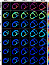
T1-weighted, Fractional Anisotropy (FA), Mean Diffusivity (MD), primary (e1), secondary (e2), and tertiary (e3) eigenvalue maps for representative short-axis slices. Infarct and border regions are visible from diffusion tensor invariants, eigenvalues, and Rd maps. Images are shown for subject 3 in the current study.
Fig. 3.
Fig. 3.
Rain-cloud plots [1] overlaid to the corresponding box plots for FA, MD, Mode, eigenvalues e1, e2, and e3, radial diffusivity (Rd), normalized T1- and T2-weighted signal intensities in the infarct, border, and remote myocardial regions.
Fig.4.
Fig.4.
Bar plots illustrating percent differences in median FA, MD, Mode, e1, e2, e3, Rd, T1-weighted, and T2-weighted normalized signal intensities in the border and infarct regions with respect to the remote myocardium. The dotted line indicates the median percentage change when data from all subjects are considered together. The percent differences reported directly on the plots are computed between remote-border and remote-infarct overall medians.

References

    1. Allen M, Poggiali D, Whitaker K, Marshall TR, Kievit RA: Raincloud plots: a multi-platform tool for robust data visualization. Wellcome open research 4 (2019) - PMC - PubMed
    1. Chen J, Song SK, Liu W, McLean M, Allen JS, Tan J, Wickline SA, Yu X: Remodeling of cardiac fiber structure after infarction in rats quantified with diffusion tensor MRI. American Journal of Physiology-Heart and Circulatory Physiology 285(3), H946–H954 (2003) - PubMed
    1. Cork TE, Perotti LE, Verzhbinsky IA, Loecher M, Ennis DB: High-Resolution Ex Vivo Microstructural MRI After Restoring Ventricular Geometry via 3D Printing. In: International Conference on Functional Imaging and Modeling of the Heart. pp. 177–186. Springer; (2019) - PMC - PubMed
    1. Kung GL, Vaseghi M, Gahm JK, Shevtsov J, Garfinkel A, Shivkumar K, Ennis DB: Microstructural infarct border zone remodeling in the post-infarct swine heart measured by diffusion tensor MRI. Frontiers in physiology 9, 826 (2018) - PMC - PubMed
    1. Li X, Perotti LE, Martinez JA, Duarte-Vogel SM, Ennis DB, Wu HH: Real-time 3T MRI-guided cardiovascular catheterization in a porcine model using a glass-fiber epoxy-based guidewire. PloS one 15(2), e0229711 (2020) - PMC - PubMed

LinkOut - more resources