. 2021 Oct 27;6(5):e0070721.
doi: 10.1128/mSphere.00707-21. Epub 2021 Sep 29.

Candida albicans ENT2 Contributes to Efficient Endocytosis, Cell Wall Integrity, Filamentation, and Virulence

Affiliations

Candida albicans ENT2 Contributes to Efficient Endocytosis, Cell Wall Integrity, Filamentation, and Virulence

Christiane Rollenhagen et al. mSphere. .

Abstract

Epsins play a pivotal role in the formation of endocytic vesicles and potentially provide a linkage between endocytic and other trafficking pathways. We identified a Candida albicans epsin, ENT2, that bears homology to the Saccharomyces cerevisiae early endocytosis genes ENT1 and ENT2 and studied its functions by a reverse genetic approach utilizing CRISPR-Cas9-mediated gene deletion. The C. albicans ent2Δ/Δ null mutant displayed cell wall defects and altered antifungal drug sensitivity. To define the role of C. albicans ENT2 in endocytosis, we performed assays with the lipophilic dye FM4-64 that revealed greatly reduced uptake in the ent2Δ/Δ mutant. Next, we showed that the C. albicans ent2Δ/Δ mutant was unable to form hyphae and biofilms. Assays for virulence properties in an in vitro keratinocyte infection model demonstrated reduced damage of mammalian adhesion zippers and host cell death from the ent2Δ/Δ mutant. We conclude that C. albicans ENT2 has a role in efficient endocytosis, a process that is required for maintaining cell wall integrity, hyphal formation, and virulence-defining traits. IMPORTANCE The opportunistic fungal pathogen Candida albicans is an important cause of invasive infections in hospitalized patients and a source of considerable morbidity and mortality. Despite its clinical importance, we still need to improve our ability to diagnose and treat this common pathogen. In order to support these advancements, a greater understanding of the biology of C. albicans is needed. In these studies, we are focused on the fundamental biological process of endocytosis, of which little is directly known in C. albicans. In addition to studying the function of a key gene in this process, we are examining the role of endocytosis in the virulence-related processes of filamentation, biofilm formation, and tissue invasion. These studies will provide greater insight into the role of endocytosis in causing invasive fungal infections.

Keywords: Candida albicans; biofilm; endocytosis; filamentation; membrane trafficking; pathogenesis; secretion.


Figures
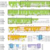
FIG 1
FIG 1
C. albicans Ent2p bears a strong homology to S. cerevisiae Ent1p/Ent2p. Shown is the alignment of S. cerevisiae (S.c) Ent1p and Ent2p with C. albicans (C.a) Ent2p, Ent3p, and Ent4p. Amino acids conserved to greater than 60% are shaded. The green boxes identify the ENTH domain. The highly conserved sequences of the ENTH domain are marked with an asterisk. The orange block delimits the pair of UIM motifs with # symbols marking the locations of conserved amino acids. Regions of low complexity and conservation have been omitted, and the extent of the omission is indicated (Δ amino acid numbers). Blue boxes indicate the tripeptide, NPF, and the purple block is the yeast clathrin-binding motif. Note that C. albicans Ent3p is presented starting from amino acid 15.
FIG 2
FIG 2
The ent2Δ null mutant strain has a modest growth defect. (A to E) The ent2Δ/Δ null mutant strain (knockout [KO]), wild type (wt), and knock-in strain (KI) were depicted in a growth curve (A), on YPD plates incubated for 48 h at 30°C (B) or 37°C (C), visualized by DIC and fluorescence microscopy stained with calcofluor white (CW) (D), as well as in a CW quantification assay (E). The bar in panel D is 5 μm. The images shown in panels B to D are representative of three different experiments. Error bars in panels A and E are the standard deviations of three different data sets. Each individual data point was run in triplicate and averaged. Statistical significance between control strains (wt and KI) and the KO strain was determined with Student’s t test (*, P < 0.05).
FIG 3
FIG 3
The ent2Δ/Δ mutant strain has delayed endocytosis and accumulates dye in the vacuole. (A) Fluorescent images of ent2Δ/Δ null mutant (KO) and control strains, wild type (wt), and knock-in (KI) strains stained with the lipophilic fluorescent dye FM4-64 are shown after incubation of 5, 15, and 30 min at room temperature (RT). (B) Quantitative analysis of the vacuolar stain in wt, KI, and KO strains after an incubation of 5, 15, and 30 min at RT. The images shown in panel A are representative of three different experiments. The bar in panel A is 5 μm. Error bars in panel B are the standard deviations from three different data sets expressed as percentage of the total number of cells.
FIG 4
FIG 4
The ent2Δ/Δ mutant strain has a decreased tolerance to cell wall stressors and salt stress. (A and C) The ent2Δ/Δ null mutant (KO), wild-type (wt), and knock-in (KI) strains were grown on YPD plates for 48 h containing cell wall stressors SDS (0.05%), calcofluor white (50 μg/ml), and Congo red (140 μg/ml) (A) as well as cationic cell stress conditions, including 500 mM KCl, 500 mM NaCl, or 100 mM LiCl (C). (B) The impact on adhesion to plastic surfaces (polystyrene) was measured using the XTT reduction assay in the ent2Δ/Δ null mutant (KO) and control strains (wt and KI) after 2 and 24 h of incubation on plastic surfaces. The images shown in panels A and C are representative of three different experiments. Error bars in panel B are the standard deviations of three different data sets. Each individual data point was run in triplicate and averaged. Statistical significance in panel B between control strains (wt and KI) and KO was determined with Student’s t test (*, P < 0.05).
FIG 5
FIG 5
The ent2Δ/Δ mutant strain has increased susceptibility to amphotericin B and caspofungin and reduced susceptibility to fluconazole. The ent2Δ/Δ null mutant (KO), wild-type (wt), and knock-in (KI) strains were grown on YPD plates for 48 h containing antifungal drugs at various concentrations as follows: amphotericin B at 0.11 μg/ml, 0.33 μg/ml, and 1.0 μg/ml (A), caspofungin at 0.025 μg/ml, 0.05 μg/ml, and 0.1 μg/ml (B), and fluconazole at 1.0 μg/ml, 2.0 μg/ml, and 4.0 μg/ml (C). The images shown in panels A to C are representative of three different experiments.
FIG 6
FIG 6
The ent2Δ/Δ mutant does not form filaments and forms an aberrant biofilm. (A) The ent2Δ/Δ null mutant (KO), wild-type (wt), and knock-in (KI) strains were assayed on agar plates grown for 72 h at 37°C with different media (medium 199 [M199] and RPMI 1640 medium) known to induce hyphal growth. (B) These strains were grown on liquid RPMI 1640 medium overnight and were visualized by DIC and fluorescence microscopy after staining with calcofluor white (CW). The bar indicates 5 μm. (C) Biofilms were grown on plastic surfaces with RPMI 1640 medium for 48 h and assayed for biofilm metabolic activity. (D) These biofilms were visualized by light microscopy using a DIC filter. The bar indicates 50 μm. The images shown in panels A, B, and D are representative of three different experiments. Error bars in panel C are the standard deviations of three different data sets. Each individual data point was run in triplicate and averaged. Statistical significance in panel C between control strains (wt and KI) and KO was determined with Student’s t test (*, P < 0.05).
FIG 7
FIG 7
The dissolution of cell-cell adhesions in human VK-2 cells is impaired in the presence of C. albicans ent2Δ/Δ. (A and B) Human VK-2 cells infected with the ent2Δ/Δ null mutant (KO), wild-type (wt), and knock-in (KI) strains for 6 and 24 h were visualized by fluorescence microscopy using an antibody against E-cadherin and DAPI for labeling the nucleus (A) and were tested by Western blotting for the proteins E-cadherin and tubulin (loading control) (B). (C) These C. albicans strains were further tested in a protease agar plate assay for their ability to lyse BSA after an overnight incubation at 30°C. The images shown in panel A to C are representative of three different experiments. The bar in panel A is 10 μm.
FIG 8
FIG 8
The C. albicans ent2Δ/Δ mutant lacks the ability to kill human VK-2 cells. VK-2 cells infected with the ent2Δ/Δ null mutant (KO), wild-type (wt), and knock-in (KI) strains for 6 h (A) and 24 h (B) were tested in a Live/Dead assay. Error bars in panels A and B are the standard deviations of three different data sets. Each individual data point was run in triplicate and averaged. Statistical significance between control strains (wt and KI) and KO was determined with Student’s t test (*, P < 0.05).

References

    1. Lee SA, Jones J, Khalique Z, Kot J, Alba M, Bernardo S, Seghal A, Wong B. 2007. A functional analysis of the Candida albicans homolog of Saccharomyces cerevisiae VPS4. FEMS Yeast Res 7:973–985. doi:10.1111/j.1567-1364.2007.00253.x. - DOI - PubMed
    1. Lee SA, Jones J, Hardison S, Kot J, Khalique Z, Bernardo SM, Lazzell A, Monteagudo C, Lopez-Ribot J. 2009. Candida albicans VPS4 is required for secretion of aspartyl proteases and in vivo virulence. Mycopathologia 167:55–63. doi:10.1007/s11046-008-9155-7. - DOI - PMC - PubMed
    1. Rane HS, Hardison S, Botelho C, Bernardo SM, Wormley F, Lee SA. 2014. Candida albicans VPS4 contributes differentially to epithelial and mucosal pathogenesis. Virulence 5:810–818. doi:10.4161/21505594.2014.956648. - DOI - PMC - PubMed
    1. Martin R, Hellwig D, Schaub Y, Bauer J, Walther A, Wendland J. 2007. Functional analysis of Candida albicans genes whose Saccharomyces cerevisiae homologues are involved in endocytosis. Yeast 24:511–522. doi:10.1002/yea.1489. - DOI - PubMed
    1. Palanisamy SKA, Ramirez MA, Lorenz M, Lee SA. 2010. Candida albicans PEP12 is required for biofilm integrity and in vivo virulence. Eukaryot Cell 9:266–277. doi:10.1128/EC.00295-09. - DOI - PMC - PubMed

MeSH terms

Substances

LinkOut - more resources