. 2021 Dec 15;85(4):e0012321.
doi: 10.1128/MMBR.00123-21. Epub 2021 Sep 29.

Sugar-Phosphate Toxicities

Affiliations

Sugar-Phosphate Toxicities

Erin F Boulanger et al. Microbiol Mol Biol Rev. .

Abstract

Accumulation of phosphorylated intermediates during cellular metabolism can have wide-ranging toxic effects on many organisms, including humans and the pathogens that infect them. These toxicities can be induced by feeding an upstream metabolite (a sugar, for instance) while simultaneously blocking the appropriate metabolic pathway with either a mutation or an enzyme inhibitor. Here, we survey the toxicities that can arise in the metabolism of glucose, galactose, fructose, fructose-asparagine, glycerol, trehalose, maltose, mannose, mannitol, arabinose, and rhamnose. Select enzymes in these metabolic pathways may serve as novel therapeutic targets. Some are conserved broadly among prokaryotes and eukaryotes (e.g., glucose and galactose) and are therefore unlikely to be viable drug targets. However, others are found only in bacteria (e.g., fructose-asparagine, rhamnose, and arabinose), and one is found in fungi but not in humans (trehalose). We discuss what is known about the mechanisms of toxicity and how resistance is achieved in order to identify the prospects and challenges associated with targeted exploitation of these pervasive metabolic vulnerabilities.

Keywords: antimicrobials; drug targets; fructose-asparagine; sugar phosphate; toxicity.


Figures
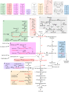
FIG 1
FIG 1
Metabolic pathways for microbial utilization of select carbohydrates. Genes whose mutation/dysfunction cause accumulations of toxic phosphosugars and the respective built-up phosphosugar are highlighted in red. Although the pathways are based on those present in E. coli, there are variations and exceptions in other bacteria. For example, the pathway shown in panel B is found in S. enterica but not in E. coli. In B. subtilis, the pgm gene and Glc-1P would be highlighted in red. No toxicities have been found in E. coli or S. enterica pathways shown in panel C, but the M. tuberculosis pathways do have toxicities (see Fig. 5). Structures for various sugars are depicted in the top panel and color coordinated with their degradation pathway in the bottom panel.
FIG 2
FIG 2
sgrR/sgrS/sgrT response to sugar-phosphate stress in E. coli. Glucose and α-methyl-glucoside (αMG) (yellow hexagons) are primarily transported by the phosphoenolpyruvate (PEP)-dependent transporter PtsG with some contribution from ManXYZ. Glucose-phosphate stress is triggered by accumulation of glucose-6-phosphate or phosphorylated αMG (yellow hexagon with attached phosphate group). During the phosphosugar stress response, SgrR induces expression of sgrS. SgrS is a noncoding RNA that base pairs with target mRNAs to either allow or protect these mRNAs from RNase E degradation, thus engendering opposite translation outcomes (upregulation, yigL; downregulation, ptsG). YigL belongs to the family of HAD-like phosphatases that can hydrolyze the phosphate group from the intoxicating sugar phosphate. SgrS also produces SgrT, which inhibits the activity of PtsG, but not ManXYZ, curtailing further transport of glucose or αMG into the cell.
FIG 3
FIG 3
Contribution of sugar phosphates to synthesis of S. enterica cell wall components. The LPS of the S. enterica galE mutant is truncated (“rough”) since UDP-galactose cannot be incorporated. In the peptidoglycan, NAM is N-acetylmuramic acid. In LPS, the rectangle symbol in represents 3-deoxy-d-manno-octulosonic acid (KDO), and the white ovals depict heptose. The orange circles in Lipid A are N-acetylglucosamine, which is derived from UDP-GlcNAc. The colors for fucose, galactose, glucose, glucuronic acid (GlcA), mannose, and N-acetylglucosamine are shown in the pathways (top) and used likewise in the peptidoglycan, LPS, and colanic acid (bottom).
FIG 4
FIG 4
LPS and peptidoglycan of a S. enterica galE mutant with various concentrations of galactose. A galE mutant of S. enterica has truncated (“rough”) LPS unless galactose is provided. Immune responses to rough LPS do not provide protection against wild-type Salmonella and are referred to as nonprotective in the figure. Trace amounts of galactose may allow some of the LPS to be full length and elicit a protective immune response, while the bacterium remains attenuated due to the regions of rough LPS still being accessible to complement and other innate immune defenses. The Goldilocks amount of galactose will restore a complete, full length, LPS, providing full virulence. Too much galactose can lead to galactose-induced toxicity, which includes accumulation of UDP-Gal and Gal-1P, and a decline in UDP-GlcNAc that ultimately leads to peptidoglycan defects and lysis.
FIG 5
FIG 5
Trehalose/maltose pathways of M. tuberculosis. Genes whose mutation/dysfunction cause accumulations of toxic phosphosugars and the respective built-up phosphosugar are highlighted in red. The E. coli versions of these pathways are shown in Fig. 1C for comparison.
FIG 6
FIG 6
Arabinose utilization and pentose phosphate pathways in E. coli. Genes whose mutation/dysfunction cause accumulations of toxic phosphosugars and the respective built-up phosphosugar are highlighted in red.

References

    1. Lee SJ, Trostel A, Adhya S. 2014. Metabolite changes signal genetic regulatory mechanisms for robust cell behavior. mBio 5:e00972-13. 10.1128/mBio.00972-13. - DOI - PMC - PubMed
    1. Oberhaensli RD, Rajagopalan B, Taylor DJ, Radda GK, Collins JE, Leonard JV, Schwarz H, Herschkowitz N. 1987. Study of hereditary fructose intolerance by use of 31P magnetic resonance spectroscopy. Lancet 2:931–934. 10.1016/s0140-6736(87)91419-x. - DOI - PubMed
    1. Yarmolinsky MB, Wiesmeyer H, Kalckar HM, Jordan E. 1959. Hereditary defects in galactose metabolism in Escherichia coli mutants, II. Galactose-induced sensitivity. Proc Natl Acad Sci USA 45:1786–1791. 10.1073/pnas.45.12.1786. - DOI - PMC - PubMed
    1. Fukasawa T, Nikaido H. 1959. Galactose-sensitive mutants of Salmonella. Nature 184(Suppl 15):1168–1169. 10.1038/1841168a0. - DOI - PubMed
    1. Fukasawa T, Nikaido H. 1959. Formation of ‘protoplasts’ in mutant strains of Salmonella induced by galactose. Nature 183:1131–1132. 10.1038/1831131a0. - DOI - PubMed

Publication types

LinkOut - more resources