. 2021 Oct 31;9(2):e0073421.
doi: 10.1128/Spectrum.00734-21. Epub 2021 Sep 29.

PGRMC1 Exerts Its Function of Anti-Influenza Virus in the Central Nervous System

Affiliations

PGRMC1 Exerts Its Function of Anti-Influenza Virus in the Central Nervous System

Kun Huang et al. Microbiol Spectr. .

Abstract

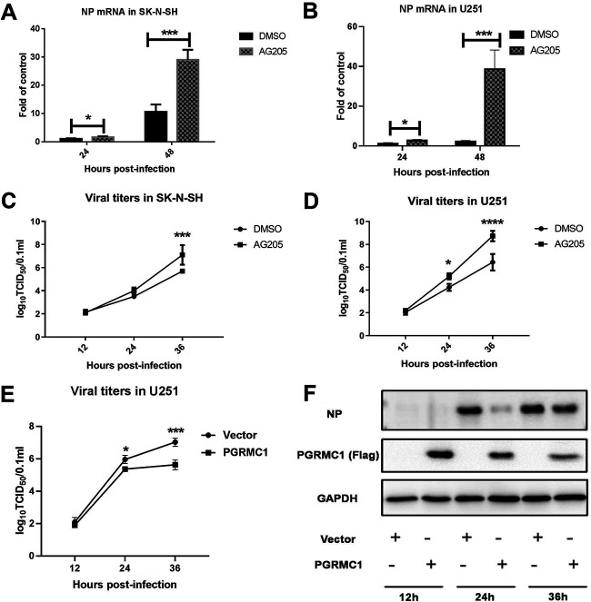
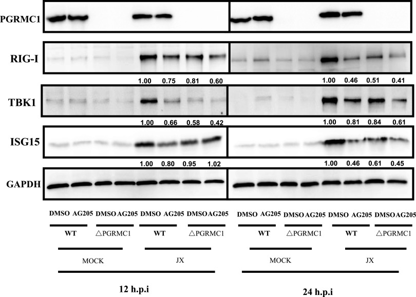
The influenza A virus (IAV) infection is usually restricted to the respiratory tract and only rarely enters the central nervous system (CNS) and causes neurological symptoms. However, the roles of host factors involved in IAV infection in the CNS remain largely undetermined. Therefore, we aimed to characterize the host responses to IAV infection in the brain. We isolated a strain of IAV H5N6, which is neurotoxic and highly pathogenic to mice. High-throughput RNA sequencing (RNA-seq) revealed 240 differentially expressed genes in IAV-infected brains. Among the significantly downregulated genes, we focused on the gene encoding progesterone receptor membrane component-1 (PGRMC1) and observed that IAV H5N6 infection clearly inhibited PGRMC1 in both neuroblastoma and glioma cells. Furthermore, treatment with AG205, a PGRMC1-specific inhibitor, or PGRMC1 knockout promoted H5N6 multiplication in vitro, while overexpression of PGRMC1 resulted in opposite effects. Furthermore, AG205 treatment or PGRMC1 knockout significantly inhibited the retinoic acid-inducible gene I (RIG-I)-mediated interferon beta (IFN-β) signaling pathway and reduced the levels of several antiviral proteins (Mx1 and ISG15). In addition, PGRMC1-mediated regulation of IFN signaling relied on inhibition of the expression and ubiquitination of RIG-I. The loss of PGRMC1 leads to an increased susceptibility of mice (brain and lung) to influenza A virus infection. Conclusively, our results show for the first time that IAV H5N6 downregulates PGRMC1 expression to contribute to virus proliferation by inhibiting RIG-I-mediated IFN-β production in the brain. These findings may offer new insights regarding the interplay between IAV and host factors that may impact IAV pathogenicity in the brain. IMPORTANCE Central nervous system (CNS) disease is one of the most common extra-respiratory tract complications of influenza A virus (IAV) infections. However, there is still little knowledge about IAV regulating host responses in brain. In this study, we identified progesterone receptor membrane component-1 (PGRMC1) as a novel host factor involved in the replication and propagation of IAV H5N6 in the host brain. We also observed that PGRMC1 antagonism was required for viral evasion from the host immune response during IAV infection via inhibition of the retinoic acid-inducible gene I (RIG-I)-mediated interferon beta (IFN-β) signaling pathway and downstream antiviral gene expression. This study revealed a newly identified regulatory mechanism used by IAV H5N6 to ensure its life cycle in the CNS.

Keywords: CNS; H5N6 virus; PGRMC1; RIG-I; virus replication.


Figures

FIG 1
FIG 1
Infection of BALB/c mice (n = 11) with H5N6 virus. Mice receiving PBS were used as controls. The mice were monitored for 14 days. (A) Body weight changes were depicted as the percentage of the starting weight of mice (*, P < 0.05; **, P < 0.01; ***, P < 0.001; ****, P < 0.0001; using two-way ANOVA). (B) The mice developed neurological symptoms. (C) Survival of infected mice (Mantel-Cox log-rank test). (D) Infectious virion titers in lung and brain were calculated through TCID50. (E) Immunohistochemical analysis of the nucleoprotein antigen in brainstem. (F to H) Degeneration and necrosis of neurons and local glial cell nodules in the cerebral cortex. The data shown are representative of three independent experiments with similar results.
FIG 2
FIG 2
Analysis of the transcriptome profiles of brain tissue after infection with H5N6 virus. (A) Gene volcano plot. (B) Heat map. (C) Scatterplot showing Gene Ontology (GO) and (D) Kyoto Encyclopedia of Genes and Genomes (KEGG) enrichment analysis.
FIG 3
FIG 3
JX inhibited PGRMC1 expression in vitro. PGRMC1 expression level in SK-N-SH cells (A and C) and U251 cells (B and D). Cells were infected with JX at an MOI of 0.01. Samples were collected at 24 and 48 hpi, followed by qRT-PCR and Western blotting to determine the mRNA (A and B) and protein (C and D) levels of PGRMC1. For real-time PCR analysis, the mRNA level was normalized to the GAPDH level. For Western blotting, the band intensities were analyzed using ImageJ, and GAPDH was used as a control in each time point. The relative PGRMC1 levels (PGRMC1/GAPDH) are shown. The data are presented as the mean ± SD from three independent experiments (*, P < 0.05; **, P < 0.01; ***, P < 0.001; ****, P < 0.0001; using two-way ANOVA).
FIG 4
FIG 4
Specific inhibition of PGRMC1 by AG205 increased JX H5N6 virus replication. SK-N-SH cells (A and C) and U251 cells (B and D) were pretreated with 15 μM AG205 or DMSO over 24 h. Cells were infected with JX at an MOI of 0.01. Samples were collected at different time points, and viral NP mRNA and viral titers were determined using qRT-PCR and TCID50, respectively. U251 cells (E and F) were transfected with Flag-PGRMC1 and empty vectors. After 24 h, the cells were infected with JX at MOI of 0.01 and expression was detected using Western blotting. TCID50 assays (E) for the virus titer of JX were performed to estimate virus multiplication at 12, 24, and 36 hpi. The expressions of PGRMC1 and H5N6 NP protein in U251 cells were detected by Western blot (F). The data are presented as the mean ± SD from three independent experiments (*, P < 0.05; **, P < 0.01; ***, P < 0.001; ****, P < 0.0001; using two-way ANOVA).
FIG 5
FIG 5
AG205 negatively modulated the IFN-β-inducing pathway. U251 cells were pretreated with 15 μM AG205 or DMSO for 24 h. The cells were infected with JX at an MOI of 0.01 (A) or were stimulated with 100 ng of poly (I·C) (B to F) for 24 h. The mRNA levels of the target genes were detected and normalized to the GAPDH level, and the expression in the DMSO and poly (I·C)-unstimulated groups were set to 1. U251 cells were transfected with Flag-PGRMC1 and empty vectors (G). After 24 h, the cells were stimulated with 100 ng of poly (I·C) for 24 h and IFN-β mRNA levels were detected. The data are presented as the mean ± SD from three independent experiments (*, P < 0.05; **, P < 0.01; ***, P < 0.001; ****, P < 0.0001; A was analyzed by repeated measures two-way ANOVA; B to G were analyzed by one-way ANOVA).
FIG 6
FIG 6
AG205 or PGRMC1 knockout inhibited the RIG-I-mediated IFN-β signaling pathway. PGRMC1-KO U251 cells and wild-type cells were treated with 15 μM AG 205 or DMSO for 24 h. Cells were infected with JX at MOI of 0.01. Samples were collected at 12 and 24 h. IFN-β signaling molecules were detected using Western blotting. The band intensities were analyzed using ImageJ, and GAPDH was used as the control.
FIG 7
FIG 7
AG205 inhibited IFN-β promoter activity induced by RIG-I-mediated IFN-β signaling molecules. Effect of AG205 or PGRMC1 on the activation of IFN-β promoter induced by RIG-I (A), MDA5 (B), IKKε (C) TBK1 (D), IRF3 (E), and IRF3-5D (F). HEK293T cells were treated with 15 μM AG 205 or DMSO for 24 h. Then, the cells were transfected with IFN-β-luc, pRL-TK, and the indicated expression plasmids of the signaling molecules. The effect of different doses of PGRMC1 (G) or AG205 (H) on the activation of IFN-β promoter induced by RIG-I was measured 24 h posttransfection using the luciferase activity assay. The expression level of each signaling molecule was detected using Western blotting with an anti-Flag antibody. The data are presented as the mean ± SD from three independent experiments (*, P < 0.05; **, P < 0.01; ***, P < 0.001; ****, P < 0.0001; A to F were analyzed by repeated measures two-way ANOVA; G and H were analyzed by one-way ANOVA).
FIG 8
FIG 8
Effect of PGRMC1 knockout on IAV replication and the RIG-I-mediated IFN-β signaling pathway. Generation of PGRMC1-KO U251 cells using the CRISPR-Cas9 system. PGRMC1 knockout was confirmed using Western blotting (A). PGRMC1-KO U251 cells (ΔPGRMC1) or wild-type U251 cells (WT) were infected with JX H5N6 virus at MOI of 0.01. Viral titers were determined using TCID50 on MDCK cells (B). The mRNA levels of the IFN-β were detected and normalized to the GAPDH level (C) (means ± SD from three independent experiments; *, P < 0.05; **, P < 0.01; ***, P < 0.001; ****, P < 0.0001; using two way ANOVA).
FIG 9
FIG 9
PGRMC1 knockout inhibited RIG-I ubiquitination. PGRMC1-KO U251 cells and wild-type cells were transfected with Flag-RIG-I and pUb-HA. After 24 h, the cells were infected with JX at an MOI of 0.01. After 24 h, the cells were subjected to anti-Flag immunoprecipitation and immunoblotting with anti-HA to monitor RIG-I ubiquitination.
FIG 10
FIG 10
PGRMC1 knockout promoted IAV replication in mice. The PGRMC1 KO mice were made by using the CRISPR-Cas9 technique (A). The analyses of protein expressions in WT and PGRMC1 KO mice by PCR (B) and Western blotting using the antibody against PGRMC1 and actin (C). C57BL/6J WT and PGRMC1 KO female mice (n = 5) aged 4 to 6 weeks were infected intranasally with 50 μl of 105 TCID50 H5N6 virus. Body weights were monitored daily for 5 days (D), and three mice in each group were euthanized at 5 dpi. IFN-β mRNA in lung (E) and viral NP mRNA in lung (F) and brain (G) were determined through qRT-PCR. Hematoxylin and eosin (H&E) staining of lung and brain sections from WT and PGRMC1 KO mice infected H5N6 virus (H). Astrocytic hyperplasia, red arrow; neuronophagia, green arrow. (I) Viral NP proteins (black arrows) were detected in the cerebral cortex of WT and PGRMC1 KO mice. Scale bars (black), 200 μm. Scale bars (red), 50 μm. Scale bars (green), 10 μm. (*, P < 0.05; **, P < 0.01; ***, P < 0.001).

Similar articles

Cited by

References

    1. Jiao P, Song H, Liu X, Song Y, Cui J, Wu S, Ye J, Qu N, Zhang T, Liao M. 2016. Pathogenicity, transmission and antigenic variation of H5N1 highly pathogenic avian influenza viruses. Front Microbiol 7:635. doi:10.3389/fmicb.2016.00635. - DOI - PMC - PubMed
    1. Ding X, Luo J, Quan L, Wu A, Jiang T. 2017. Evolutionary genotypes of influenza A (H7N9) viruses over five epidemic waves in China. Infect Genet Evol 55:269–276. doi:10.1016/j.meegid.2017.09.027. - DOI - PubMed
    1. Bi Y, Chen Q, Wang Q, Chen J, Jin T, Wong G, Quan C, Liu J, Wu J, Yin R, Zhao L, Li M, Ding Z, Zou R, Xu W, Li H, Wang H, Tian K, Fu G, Huang Y, Shestopalov A, Li S, Xu B, Yu H, Luo T, Lu L, Xu X, Luo Y, Liu Y, Shi W, Liu D, Gao GF. 2016. Genesis, evolution and prevalence of H5N6 avian influenza viruses in China. Cell Host Microbe 20:810–821. doi:10.1016/j.chom.2016.10.022. - DOI - PubMed
    1. You F, Sun H, Zhou X, Sun W, Liang S, Zhai Z, Jiang Z. 2009. PCBP2 mediates degradation of the adaptor MAVS via the HECT ubiquitin ligase AIP4. Nat Immunol 10:1300–1308. doi:10.1038/ni.1815. - DOI - PubMed
    1. Iwasaki A, Pillai PS. 2014. Innate immunity to influenza virus infection. Nat Rev Immunol 14:315–328. doi:10.1038/nri3665. - DOI - PMC - PubMed

Publication types