. 2021 Nov 9;5(21):4515-4520.
doi: 10.1182/bloodadvances.2021004621.

Rational biomarker development for the early and minimally invasive monitoring of AML

Affiliations

Rational biomarker development for the early and minimally invasive monitoring of AML

Sherif Abdelhamed et al. Blood Adv. .

Abstract

Recurrent disease remains the principal cause for treatment failure in acute myeloid leukemia (AML) across age groups. Reliable biomarkers of AML relapse risk and disease burden have been problematic, as symptoms appear late and current monitoring relies on invasive and cost-ineffective serial bone marrow (BM) surveillance. In this report, we discover a set of unique microRNA (miRNA) that circulates in AML-derived vesicles in the peripheral blood ahead of the general dissemination of leukemic blasts and symptomatic BM failure. Next-generation sequencing of extracellular vesicle-contained small RNA in 12 AML patients and 12 controls allowed us to identify a panel of differentially incorporated miRNA. Proof-of-concept studies using a murine model and patient-derived xenografts demonstrate the feasibility of developing miR-1246, as a potential minimally invasive AML biomarker.


Figures

Figure 1.
Figure 1.
Correlation between EVs levels and AML disease burden. (A) In vivo imaging of mice xenografted with Molm-14 cells expressing luciferase at 1, 2, and 3 weeks. Images were taken by IVIS (left), and values were normalized to controls to calculate fold change (right). (B) Correlative analysis of tumor burden between human CD45 (hCD45) levels measured by flow cytometer in both the PB and BM in red vs luciferase activity in gray. (C) EV quantification using NanoSight analyzer from Molm-14 xenografted (red) or control (black) mice. Data show relative vesicles abundance over time (left) and correlation analysis at week 3 of PB and BM (red) vs EV abundance (blue). (D) Cryo-TEM image of Molm-14–derived EVs. Scale bar is 100 nm. (E) Workflow for measuring circulating EVs in the PB of mice bearing mGFP+ AML cells. (F) EV counts per unit volume (1 × 10⁻⁴ μL) correspond to BM tumor burden. (G) Quantification of relative AML EV count in the PB compared with total lipid vesicles in plasma. Each point represents EV count per unit volume (1 × 10⁻⁴ μL). (H) Representative 3-dimensional images and concentration of 20% (top) and 50% (bottom) animal cohorts. Bounding box represents 100 μm × 100 μm × 10 μm volume. Concentrations determined using [EVs/µL = (average EV count) × (volume/1) × (dilution factor)]. mGFP (white). (I) PB plasma of AML xenografted NSG mice contain numerous lipid vesicles and a discrete mGFP EV population (right) not seen in nonengrafted control animals (left). CellMask Lipid Dye (red), mGFP (green). Bounding box is 5 μm x 5 μm. Images captured with CoreDV epifluorescence microscope with 100× 1.49 TIRF objective and Nikon Coolpix CCD camera. Significance determined by Student t test. *P < .05; ***P < .001.
Figure 2.
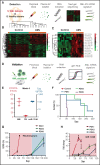
Figure 2.
Identification of AML miRNA signature from patients’ plasma EVs . (A) Workflow for characterization of EVs miRNA content. The RNA content of EVs from blood plasma collected from 12 AML patients and 12 healthy donors were sequenced using Nextseq500 analyzer. (B) Heat map showing the 243 differentially expressed miRNA between the AML patients (orange) and the healthy donors (green) groups. (C) Heat map showing the AML signature composed of 15 miRNAs uniquely upregulated in the AML patients (orange) vs the healthy donors (green). (D) Workflow for validation of miR-1246, a candidate miRNA among the AML signature. Molm-14 cells or PDXs were executed, and miR-1246 levels were measured in the blood plasma EVs by quantitative polymerase chain reaction (qPCR). (E) Correlative analysis at 4 weeks in Molm-14 xenografted mice between human CD45 levels measured by flow cytometer in both the PB and BM (red), and miR-1246 levels measured by qPCR and normalized to U6 control (blue). (F) Survival curve of PDXs (n = 5 per group; PDX1, PDX2, and PDX5). (G) Flow cytometer analysis showing PB chimerism represented by hCD45 levels over time in mice (n = 5 per group) xenografted with PDX1, PDX2, and PDX5. (H) qPCR analysis showing fold change of miR-1246 over time in PDX1 (red), PDX2 (blue), and PDX5 (green) relative to week 2. Data were normalized to U6 control (dCT), and fold change was determined relative to nonengrafted controls (ddCT).

References

    1. Loken MR, Alonzo TA, Pardo L, et al. . Residual disease detected by multidimensional flow cytometry signifies high relapse risk in patients with de novo acute myeloid leukemia: a report from Children’s Oncology Group. Blood. 2012;120(8):1581-1588. - PMC - PubMed
    1. Yin JA, O’Brien MA, Hills RK, Daly SB, Wheatley K, Burnett AK.. Minimal residual disease monitoring by quantitative RT-PCR in core binding factor AML allows risk stratification and predicts relapse: results of the United Kingdom MRC AML-15 trial. Blood. 2012;120(14):2826-2835. - PubMed
    1. Döhner H, Estey EH, Amadori S, et al. ; European LeukemiaNet . Diagnosis and management of acute myeloid leukemia in adults: recommendations from an international expert panel, on behalf of the European LeukemiaNet. Blood. 2010;115(3):453-474. - PubMed
    1. Hageman IM, Peek AM, de Haas V, Damen-Korbijn CM, Kaspers GJ.. Value of routine bone marrow examination in pediatric acute myeloid leukemia (AML): a study of the Dutch Childhood Oncology Group (DCOG). Pediatr Blood Cancer. 2012;59(7):1239-1244. - PubMed
    1. Hornick NI, Huan J, Doron B, et al. . Serum exosome microRNA as a minimally-invasive early biomarker of AML. Sci Rep. 2015;5(1):11295. - PMC - PubMed

Publication types