. 2021 Dec;36(1):2160-2169.
doi: 10.1080/14756366.2021.1983808.

Development of a fluorogenic ADAMTS-7 substrate

Affiliations

Development of a fluorogenic ADAMTS-7 substrate

Salvatore Santamaria et al. J Enzyme Inhib Med Chem. 2021 Dec.

Abstract

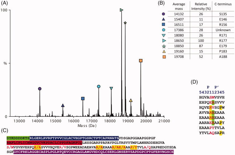
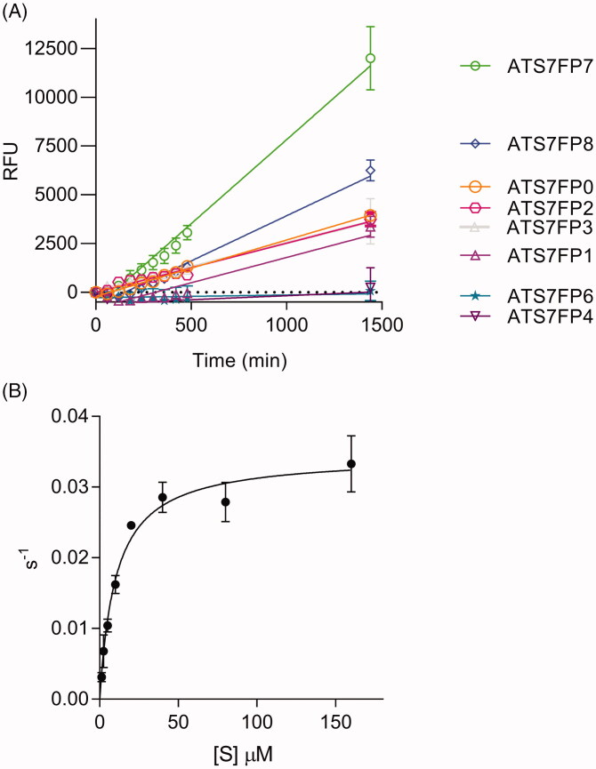
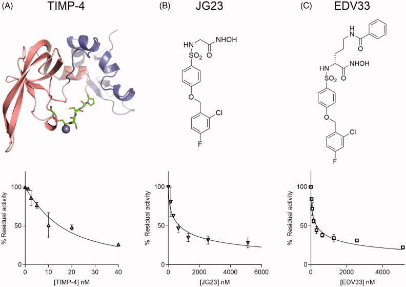
The extracellular protease ADAMTS-7 has been identified as a potential therapeutic target in atherosclerosis and associated diseases such as coronary artery disease (CAD). However, ADAMTS-7 inhibitors have not been reported so far. Screening of inhibitors has been hindered by the lack of a suitable peptide substrate and, consequently, a convenient activity assay. Here we describe the first fluorescence resonance energy transfer (FRET) substrate for ADAMTS-7, ATS7FP7. ATS7FP7 was used to measure inhibition constants for the endogenous ADAMTS-7 inhibitor, TIMP-4, as well as two hydroxamate-based zinc chelating inhibitors. These inhibition constants match well with IC50 values obtained with our SDS-PAGE assay that uses the N-terminal fragment of latent TGF-β-binding protein 4 (LTBP4S-A) as a substrate. Our novel fluorogenic substrate ATS7FP7 is suitable for high throughput screening of ADAMTS-7 inhibitors, thus accelerating translational studies aiming at inhibition of ADAMTS-7 as a novel treatment for cardiovascular diseases such as atherosclerosis and CAD.

Keywords: ADAMTS-7; ADAMTS7; activity assay; coronary artery disease; inhibitor.


Conflict of interest statement

No potential conflict of interest was reported by the author(s).

Figures

Figure 1.
Figure 1.
Mass spectrometry of the recombinant protein construct LTBP4S-A cleaved by ADAMTS-7. (A) Mass spectra of LTBP4S-A cleavage fragments showing the average mass in Dalton (Da) and relative peak intensities as a percentage of the highest peak. Cysteine residues were oxidised (B) Eight out of nine peaks shown could be matched to a cleavage fragment. The identified fragments all start with Asp1 of the N-terminal FLAG tag and have a C-terminus generated by ADAMTS-7 cleavage (P1 of the cleavage site). The relative peak intensities are an indication of the relative amounts best described as semi-quantitative. (C) Amino acid sequence of the recombinant protein construct LTBP4S-A. The residues of which the peptide bonds were cleaved by ADAMTS-7 are shown in red. Cleavage sites that we identified previously using a different method are highlighted in yellow. The N-terminal FLAG tag is highlighted in green, the 4-cys domain in blue, the EGF-like domain in red and the hybrid domain in purple. (D) Cleavage sites in LTBP4S-A established in this study. The amino acid sequences surrounding the scissile bonds identified in this study by native MS are shown. The amino acids in P1 and P1′ that form the scissile bond are shown in red. Arginine residues in P1 and proline residues in P4′ are highlighted in yellow. Alanine residues in P1’ are highlighted in green.
Figure 2.
Figure 2.
ATS7FP7 is cleaved efficiently by ADAMTS-7. (A) Cleavage of FRET peptides based on LTBP4 by ADAMTS-7. FRET peptides (40 μM) were incubated with ADAMTS-7 (10 nM). Fluorescence was detected (λex = 485 nm, λem = 520 nm) for 24 h at 37 °C and reported as relative fluorescence units (RFU). The data are presented as average ± SEM (n = 3) and were fitted to a linear regression using Graphpad Prism (B) Cleavage of FRET peptide ATS7FP7 by ADAMTS-7 (10 nM) is shown. Data were fitted to the Michaelis–Menten equation and are presented as average ± SEM (n = 3).
Figure 3.
Figure 3.
Inhibition of ADAMTS-7 cleavage of ATS7FP7 by TIMP-4, JG23, and EDV33. ADAMTS-7 was incubated for 1 h either at 5 nM with TIMP-4 (A), JG23 (B) or EDV33 (C) at 10 nM before addition of ATS7FP7 (40 μM). Percent of inhibition was calculated from control reactions containing only DMSO. For TIMP-4, data were fitted to the Morrison equation, while in case of JG23 or EDV33 the IC50 equation was used. The data are presented as average ± SEM (n = 3). Structures of TIMP-4, JG23 and EDV33 are shown above the graphs. In (A), the model of TIMP-4 was generated using homology modelling with HHpred and MODELLER. The N-terminal domain is shown in pink, the C-terminal domain in blue. Residues that sit in the active site of metalloproteases are shown as sticks with carbon in green. From left to right: Cys102 disulphide bonded to Cys30 (first residue in the mature protein), Ser31-Pro34. Chelated Zinc (Zn2+) in the active site of metalloproteases is shown as a blue sphere.
Scheme 1.
Scheme 1.
Synthesis of EDV33. Reagents and conditions: i) (R)-N-Boc-ornithine, Et3N, 1:1 H2O-Dioxane, 18 h (99.6%); ii) THPONH2, HOBT, NMM, EDC, DMF, 18 h (49.2%); iii) TFA, CH2Cl2, 0 °C, 30 min. (40.7%); iv) benzoyl chloride, DIPEA, DMF, 18 h (64.8%); v) 4 N HCl, dioxane, MeOH, 1.5 h (71.5%).
Figure 4.
Figure 4.
Inhibition of ADAMTS-7 cleavage of LTBP4S-A by JG23, and EDV33. ADAMTS-7 (19 nM) was incubated for 2 h with various concentrations of JG23 (A) or EDV33 (B) before addition of LTBP4S-A (5.6 μM). After 17 h, proteolysis was stopped by addition of EDTA and monitored by densitometry following SDS-PAGE/Coomassie Brilliant Blue staining. Representative gels are shown. The data in the plots are presented as average ± SEM (n = 3) and were fitted to nonlinear regression analysis for determination of IC50 values.
Figure 5.
Figure 5.
Result of the molecular dynamics simulation and proposed binding mode of EDV33 into the ADAMTS-7 metalloproteinase domain. (A) Homology model of the ADAMTS-7 metalloprotease domain (grey) and disintegrin-like domain (pink). The hydroxamate group binds to the zinc ion (Zn2+) in the active site (blue sphere). EDV33 is shown with green carbon atoms. (B) Zoom in of the active site. The surface of the active site is shown in a transparent representation, showing EDV33 and key interacting residues. ADAMTS-7 residues are shown with grey carbon atoms, red oxygen atoms and blue nitrogen atoms. The amino acid numbering starts from the first methionine in the signal peptide (UniProt ID Q9UKP4). For molecular dynamics simulation, see also Supplementary Figures 4 and 5.

References

    1. Pu X, Xiao Q, Kiechl S, et al. . ADAMTS7 cleavage and vascular smooth muscle cell migration is affected by a coronary-artery-disease-associated variant. Am J Hum Genet 2013;92:366–74. - PMC - PubMed
    1. Mizoguchi T, MacDonald BT, Bhandary B, et al. . Coronary disease association with ADAMTS7 is due to protease activity. Circ Res 2021;129.4:458–70. - PMC - PubMed
    1. Reilly MP, Li M, He J, Ferguson JF, et al. . Identification of ADAMTS7 as a novel locus for coronary atherosclerosis and association of ABO with myocardial infarction in the presence of coronary atherosclerosis: two genome-wide association studies. Lancet 2011;377:383–92. - PMC - PubMed
    1. Schunkert H, König IR, Kathiresan S, et al. . Large-scale association analysis identifies 13 new susceptibility loci for coronary artery disease. Nat Genet 2011;43:333–8. - PMC - PubMed
    1. Bengtsson E, Hultman K, Dunér P, et al. . ADAMTS-7 is associated with a high-risk plaque phenotype in human atherosclerosis. Sci Rep 2017;7:3753. - PMC - PubMed

MeSH terms