. 2021 Sep 29;21(1):184.
doi: 10.1186/s12862-021-01912-8.

A new extinct species of alligator lizard (Squamata: Elgaria) and an expanded perspective on the osteology and phylogeny of Gerrhonotinae

Affiliations

A new extinct species of alligator lizard (Squamata: Elgaria) and an expanded perspective on the osteology and phylogeny of Gerrhonotinae

Simon G Scarpetta et al. BMC Ecol Evol. .

Abstract

Background: Alligator lizards (Gerrhonotinae) are a well-known group of extant North American lizard. Although many fossils were previously referred to Gerrhonotinae, most of those fossils are isolated and fragmentary cranial elements that could not be placed in a precise phylogenetic context, and only a handful of known fossils are articulated skulls. The fossil record has provided limited information on the biogeography and phylogeny of Gerrhonotinae.

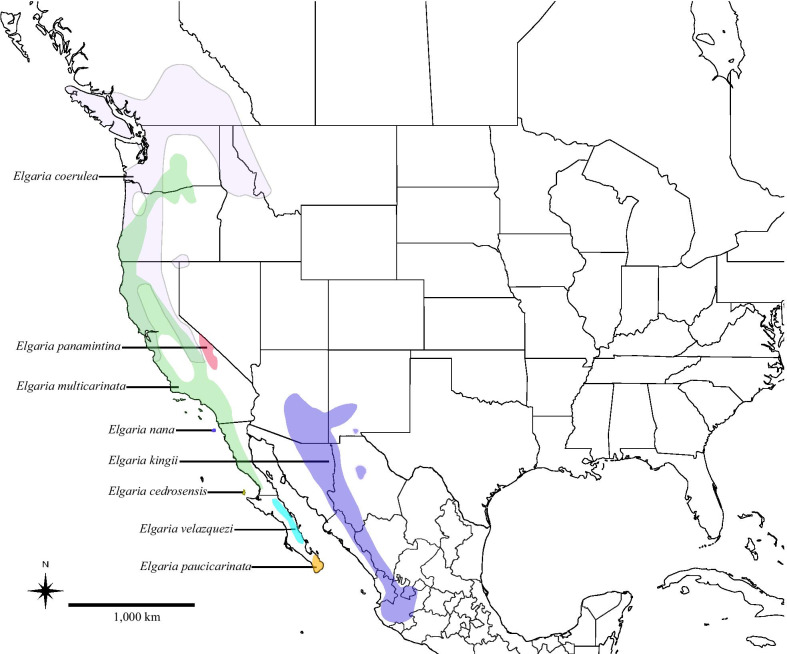
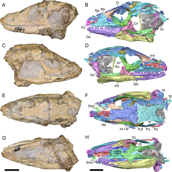
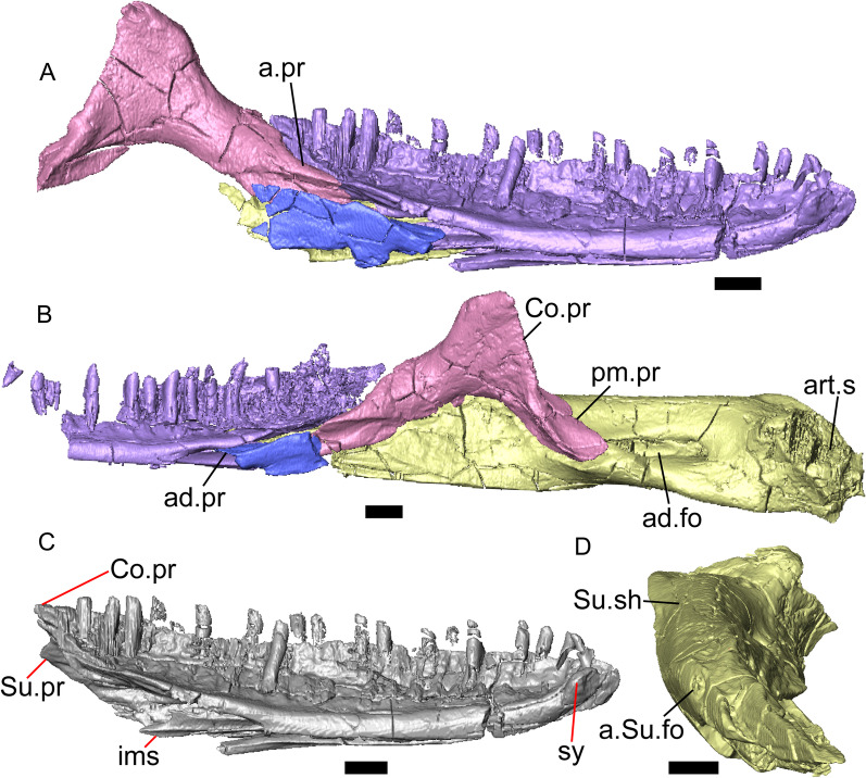
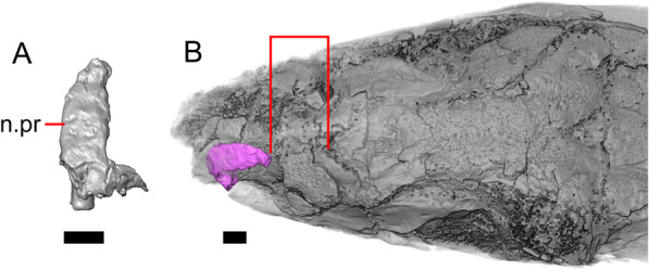
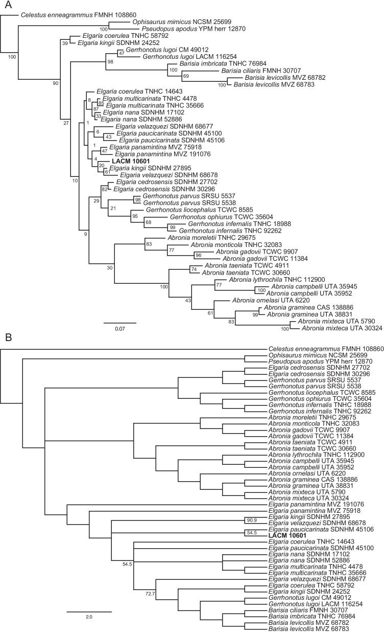
Results: We redescribe a nearly complete articulated fossil skull from the Pliocene sediments of the Anza-Borrego Desert in southern California, and refer the specimen to the alligator lizard genus Elgaria. The fossil is a representative of a newly described species, Elgaria peludoverde. We created a morphological matrix to assess the phylogeny of alligator lizards and facilitate identifications of fossil gerrhonotines. The matrix contains a considerably expanded taxonomic sample relative to previous morphological studies of gerrhonotines, and we sampled two specimens for many species to partially account for intraspecific variation. Specimen-based phylogenetic analyses of our dataset using Bayesian inference and parsimony inferred that Elgaria peludoverde is part of crown Elgaria. The new species is potentially related to the extant species Elgaria kingii and Elgaria paucicarinata, but that relationship was not strongly supported, probably because of extensive variation among Elgaria. We explored several alternative biogeographic scenarios implied by the geographic and temporal occurrence of the new species and its potential phylogenetic placements.

Conclusions: Elgaria peludoverde is the first described extinct species of Elgaria and provides new information on the biogeographic history and diversification of Elgaria. Our research expands the understanding of phylogenetic relationships and biogeography of alligator lizards and strengthens the foundation of future investigations. The osteological data and phylogenetic matrix that we provided will be critical for future efforts to place fossil gerrhonotines. Despite limited intraspecific sampled sizes, we encountered substantial variation among gerrhonotines, demonstrating the value of exploring patterns of variation for morphological phylogenetics and for the phylogenetic placement of fossils. Future osteological investigations on the species we examined and on species we did not examine will continue to augment our knowledge of patterns of variation in alligator lizards and aid in phylogenetics and fossil placement.

Keywords: Anza-Borrego; Biogeography; Elgaria; Fossils; Gerrhonotinae; Morphology; Osteology; Phylogenetics; Pliocene.


Conflict of interest statement

The authors declare that they have no competing interests.

Figures

Fig. 1
Fig. 1
Geographic range distributions of modern species of Elgaria
Fig. 2
Fig. 2
Late Neogene and early Pleistocene temporal framework showing correlation of known chrons with absolute time
Fig. 3
Fig. 3
Holotype of E. peludoverde (LACM 10601). Images on the left column are of the physical specimen, and those on the right column are surface renderings of the segmented fossil. Scale bars = 5 mm. a, b Skull in left lateral view. c, d Skull in right lateral view. e, f Skull in dorsal view. g, h Skull in ventral view. bc braincase, Co coronoid, De dentary, Ec ectopterygoid, Ep epipterygoid, Fr Frontal, Ju jugal, md mandible, Mx maxilla, os osteoderm, os.l.fp osteoderm overlying the frontoparietal shield, Na nasal, P parietal, Pa palatine, Po postorbital, Pof postfrontal, Prf prefrontal, Pt pterygoid, Px premaxilla, Qu quadrate, Smx septomaxilla, Spl splenial, St supratemporal, Sq squamosal, Vo vomer
Fig. 4
Fig. 4
Mandibular elements of LACM 10601. Scale bars = 1 mm. a Left mandible in medial view. b Right mandible in medial view. c Left dentary in medial view. d Right surangular in anterior view. a.pr anterior process, a.Su.fo anterior surangular foramen, ad.fo adductor fossa, ad.pr anterodorsal process, art.s articular surface, Co.pr coronoid process (of either the coronoid or the dentary), ims intramandibular septum, pm.pr posteromedial process, Su.pr surangular process, Su.sh surangular shelf, sy symphysis
Fig. 5
Fig. 5
Select cranial elements of LACM 10601. Scale bars = 1 mm. a Left maxilla in medial view. b Left maxilla in anterior view. c Septomaxilla fragments. d Left nasal fragment in ventral view. e Vomers in posterior view. a.pr anterior process, f.pr facial process, mx.lp maxillary lappet, or.pr orbital process, p.pr posterior process, pd.fl posterodorsal flange, Px.pr premaxillary process
Fig. 6
Fig. 6
Palatal elements of LACM 10601. Scale bars = 1 mm. a Left ectopterygoid in ventral view. b Palatines in ventral view. c Pterygoids in dorsal view. d Pterygoids in ventral view. ch.gr choanal groove, d.pr dorsal process, d.r dorsal ridge, mx.ft maxillary facet, mx.pr maxillary process, p.Ep.gr postepipterygoid groove, pa.pl palatal plate, pd.r posterodorsal ridge, Pr.fl pterygoid flange, Pt.te pterygoid teeth, Q.pr quadrate process, v.pr ventral process, v.r ventral ridge
Fig. 7
Fig. 7
Premaxilla and spatial position of the premaxilla with respect to the frontal of LACM 10601. Scale bars = 1 mm. a premaxilla in anterior view. b Partially transparent volume rendering of the anterior portion of the skull, and (in magenta) a surface rendering of the premaxilla transformed to be at its natural position (using the transform module in Avizo Lite 9). Red lines mark the anterior margin of the frontal and the posterior margin of the premaxilla. n.pr nasal process
Fig. 8
Fig. 8
Select cranial elements of LACM 10601. Scale bars = 1 mm except for h, for which the scale bar = 0.5 mm. a Left prefrontal in lateral view. b Left jugal in ventrolateral view. c Left postfrontal in dorsal view. d Left postorbital in dorsal view. e Parietal in posterior view. f Right supratemporal in dorsal view. g Right squamosal in lateral view. h Left lacrimal in posterior view. a.pr anterior process (of either the supratemporal or the squamosal), J.pr jugal process, J.s jugal spur, m.s medial shelf, Mx.pr maxillary process, o.pr orbital process, Po.pr postorbital process, pp.pr postparietal process, Pof.pr postfrontal process, pv.pr posteroventral process, Sq.pr squamosal process, v.l ventral lamina
Fig. 9
Fig. 9
Frontal and palatines of LACM 10601 in right lateral view. Scale bar = 1 mm. cr.cr crista cranii, Fr frontal, Pa palatine, Pof.fa postfrontal facet, Prf.fa prefrontal facet
Fig. 10
Fig. 10
Right quadrate of LACM 10601. Scale bar = 1 mm. a Anterior view. b Posterior view. am.s anteromedial surface, co conch, Pt.lm pterygoid lamina, ty.cr tympanic crest
Fig. 11
Fig. 11
Braincase of LACM 10601. Scale bars = 1 mm. a Braincase in right lateral view. b Braincase in dorsal view. c Braincase in posterior view. aar anterior ampullar recess, bt.pr basipterygoid process, btb basiocciptal tubercle, cr.if crista interfenestralis, i.pro incisura prootica, oc occipital condyle, Ot otooccipital, Ot.cr otooccipital crest, Ot.d otooccipital depression, pocc paroccipital process, pr.as ascending process, Pro prootic, Pro.a.pr alar process of the prootic, Pro.cr prootic crest, Psp.pr parasphenoid process, So supraoccipital, Sp sphenoid, Sp.a.pr alar process of the sphenoid
Fig. 12
Fig. 12
Majority-rule consensus trees from the unconstrained phylogenetic analyses. For the Bayesian analysis, node values are posterior probabilities as a percent. For the parsimony analysis, node values indicate the percent of MPTs in which a given node occurred; if no value is listed, the node occurred in all MPTs. a Bayesian analysis. b Parsimony analysis
Fig. 13
Fig. 13
Majority-rule consensus trees from the partially constrained phylogenetic analyses in which G. parvus could attach anywhere. For the Bayesian analysis, node values are posterior probabilities as a percent. For the parsimony analysis, node values indicate the percent of MPTs in which a given node occurred; if no value is listed, the node occurred in all MPTs. a Bayesian analysis. b Parsimony analysis
Fig. 14
Fig. 14
Majority-rule consensus trees from the fully constrained phylogenetic analyses in which G. parvus was constrained to be the sister taxon of the keeled-scale Gerrhonotus. For the Bayesian analysis, node values are posterior probabilities as a percent. For the parsimony analysis, node values indicate the percent of MPTs in which a given node occurred; if no value is listed, the node occurred in all MPTs. a Bayesian analysis. b Parsimony analysis
Fig. 15
Fig. 15
Relationships among Elgaria (based on [5]) and showing the two alternative hypotheses of the relationships of E. peludoverde. a The Bayesian analysis in which G. parvus was constrained to be the sister taxon of the keeled-scale Gerrhonotus. b The Bayesian analysis in which G. parvus could attach anywhere. The top illustration is E. kingii (drawn from a photo with a CC BY-NC 4.0 license, see https://creativecommons.org/licenses/by-nc/4.0/, courtesy of Kory Roberts and downloaded from https://www.inaturalist.org/observations/27879246) and the bottom illustration is of E. multicarinata (drawn from a photo taken by SGS)
Fig. 16
Fig. 16
Premaxillae in anterior view. Scale bars = 1 mm. a E. kingii SDNHM 27985. b E. velazquezi SDNHM 68677. c E. coerulea TNHC 14643. d G. infernalis TNHC 18988. e B. imbricata TNHC 76984. f B levicollis MVZ 68783. g Abronia gadovii TCWC 9907. h A. graminea UTA 38831. i A. lythrochila TNHC 112900. a.fo anterior foramen, d.o dorsal ossification, n.pr nasal process, o.b ossified bridge
Fig. 17
Fig. 17
Premaxillae in anterior view illustrating the relative widths of the nasal process. The red lines illustrate where the width of the nasal process is widest for each specimen. Scale bars = 1 mm. a E. kingii SDNHM 24252. b Abronia gadovii TCWC 9907. c B. imbricata TNHC 76984
Fig. 18
Fig. 18
Premaxillae and contact (or lack thereof) between the premaxilla and the frontal. Scale bars = 1 mm. a, b, are A. graminea UTA 38831, c, d are E. paucicarinata SDNHM 45100. a Premaxilla in lateral view. b Premaxilla and frontal in dorsal view. c Premaxilla in lateral view. d Premaxilla and frontal in dorsal view. p.e posteroventral extension
Fig. 19
Fig. 19
Premaxillae of anguid lizards. Scale bars = 1 mm. a B. levicollis MVZ 68782 in posterior view. b E. coerulea TNHC 14643 in posterior view. c O. mimicus NCSM 25699 in posterior view. d O. mimicus NCSM 25699 in left lateral view. e G. infernalis TNHC 198988 in left lateral view. f E. velazquezi SDNHM 68678 in ventral view. g O. mimicus NCSM 25699 in ventral view. pa.pr palatal process, v.k ventral keel
Fig. 20
Fig. 20
Nasals and maxillae in dorsal view. Scale bars = 1 mm. a E. nana SDNHM 17102. b A. ornelasi UTA 6220. c G. lugoi LACM 116254. d B. levicollis MVZ 68782. a.pr anterior process, m.px.pr medialmost inflection of the premaxillary process of the maxilla
Fig. 21
Fig. 21
Septomaxillae. Scale bars = 1 mm. a E. kingii SDNHM 24252. b E. paucicarinata SDNHM 45106. c A. gadovii TCWC 9907. d G. lugoi LACM 116254. a.pr anterior process, p.pr posterior process
Fig. 22
Fig. 22
Maxillae and frontals. Scale bars = 1 mm. a Left maxilla of E. multicarinata TNHC 35666 in lateral view. b Left maxilla of A. mixteca UTA 30324 in lateral view. c Maxillae and frontal of E. coerulea TNHC 58792 in anterior view. d Maxillae and frontal of G. infernalis TNHC 18988 in anterior view. a.pr anterior process, Fr frontal, Mx maxilla, p.pr posterior process
Fig. 23
Fig. 23
Relative width of the facial process of the left maxilla, marked by red lines. Left prefrontal shown to emphasize the anterior margin of the lacrimal foramen. Scale bars = 1 mm. a A. monticola TNHC 32083. b E. cedrosensis SDNHM 27702. c G. lugoi LACM 116254. f.pr facial process
Fig. 24
Fig. 24
Left maxillae. Scale bars = 1 mm. a A. campbelli UTA 35945 in anterior view. b E. cedrosensis SDNHM 27702 in anterior view. c B. levicollis MVZ 68783 in anterior view. d E. multicarinata TNHC 35666 in dorsal view. e A. campbelli UTA 35945 in dorsal view. f G. lugoi LACM 116254 in dorsal view. f.pr facial process, mx.lp maxillary lappett, pa.pr palatine process
Fig. 25
Fig. 25
Right lacrimals. For a, b scale bars = 1 mm, for ce scale bars = 0.5 mm. a B. levicollis MVZ 68783 in lateral view. b E. paucicarinata SDNHM 45100 in lateral view. c G. parvus SRSU 5537 in posterior view. d B. levicollis MVZ 68783 in posterior view. e E. paucicarinata SDNHM 45100 in posterior view. m.s medial shelf
Fig. 26
Fig. 26
Left prefrontals in lateral view. Scale bars = 1 mm. a E. coerulea TNHC 14643. b. G. lugoi LACM 116254. a.pr anterior process
Fig. 27
Fig. 27
Left jugals. Scale bars = 1 mm. a E. cedrosensis SDNHM 27702 in ventrolateral view. b A. lythrochila TNHC 112900 in ventrolateral view. c B. levicollis MVZ 68782 in lateral view. d A. ornelasi UTA 6220 in lateral view. v.l ventral lamina
Fig. 28
Fig. 28
Frontal of P. apodus YPM 12870. Scale bars = 1 mm. a Dorsal view. b Ventral view
Fig. 29
Fig. 29
Frontals in dorsal view illustrating length and width measurements (red lines). Scale bars = 1 mm. a A. campbelli UTA 35945. b E. cedrosensis SDNHM 27702. c B. levicollis MVZ 68782
Fig. 30
Fig. 30
Parietals, supratemporals, and squamosals. Scale bars = 1 mm. a Parietal of E. paucicarinata SDNHM 45100 in posterior view. b Parietal of E. cedrosensis SDNHM 30296 in posterior view. c Right supratemporal of E. velazquezi SDNHM 68678 in dorsal view. d Right supratemporal of A. campbelli UTA 35945 in dorsal view. e Left squamosal of E. multicarinata TNHC 35666 in lateral view. f Left squamosal of A. mixteca UTA 30324 in lateral view. a.pr anterior process, p.pr posterior process, p.r posterior recess
Fig. 31
Fig. 31
Vomers. Scale bars = 1 mm. a Right vomer of A. campbelli UTA 35945 in lateral view. b Right vomer of G. infernalis TNHC 18988 in lateral view. c Right vomer of A. monticola TNHC 32083 in lateral view. d Vomers of E. paucicarinata SDNHM 45100 in dorsal view. e Vomers of A. taeniata TCWC 4911 in dorsal view. f Vomers of E. velazquezi SDNHM 68678 in posterior view. g Vomers of G. parvus SRSU 5538 in posterior view. h Vomers of A. monticola TNHC 32083 in posterior view. fo.mpn foramen for the medial palatine nerve, pd.fl posterodorsal flange, vn vomeronasal region
Fig. 32
Fig. 32
Palatine, lacrimal, jugal, and prefrontal in articulation and in dorsal view. Scale bars = 1 mm. a E. multicarinata TNHC 35666. b E. paucicarinata SDNHM 45106. c E. kingii SDNHM 24252. Ju jugal, La lacrimal, Pa Palatine, Prf prefrontal
Fig. 33
Fig. 33
Palatines and pterygoids in articulation in ventral view. Scale bars = 1 mm. a E. cedrosensis SDNHM 27702. b G. lugoi LACM 116254. c A. campbelli UTA 35952. d A. mixteca UTA 30324. e P. apodus YPM 12870
Fig. 34
Fig. 34
Palatines. Scale bars = 1 mm. a Left palatine of E. panamintina MVZ 75918 in anteroventral view (but figure oriented such that anterior is facing left). b Left palatine of A. ornelasi UTA 6220 in anteroventral view (but figure oriented such that anterior is facing left). c Left palatine of A. monticola TNHC 32083 in lateral view. d Left palatine of A. ornelasi UTA 6220 in lateral view. e Palatines of E. panamintina MVZ 75918 in anterior view. f Palatines of B. levicollis MVZ 68782 in anterior view. do.fl dorsal flange, vl.pr ventrolateral plate, v.r ventral ridge
Fig. 35
Fig. 35
Pterygoids. Scale bars = 1 mm. a Right pterygoid of A. campbelli UTA 35945 in posterodorsal view. b Right pterygoid of E. cedrosensis SDNHM 27702 in posterodorsal view. c Right pterygoid of P. apodus YPM 12870 in posterodorsal view. d Pterygoids of E. kingii SDNHM 24252 in dorsal view (anterior is facing down). e Pterygoids of E. paucicarinata SDNHM 45106 in dorsal view (anterior is facing down). d.r dorsal ridge, p.Ep.gr postepipterygoid groove, pd.r posterodorsal ridge
Fig. 36
Fig. 36
Right quadrates. Scale bars = 1 mm. a E. paucicarinata SDNHM 45100 in posterior view. b A. campbelli UTA 35952 in posterior view. c P. apodus YPM 12870 in posterior view. d E. paucicarinata SDNHM 45100 in ventral view. e A. campbelli UTA 35952 in ventral view. am.s anteromedial surface, co conch
Fig. 37
Fig. 37
Supraoccipitals, sphenoids, and left prootics (individual braincase elements). Scale bars = 1 mm. a Supraoccipital of E. coerulea TNHC 14643 in dorsal view. b Supraoccipital of G. infernalis TNHC 18988 in dorsal view. c Sphenoid of E. coerulea TNHC 14643 in anterior view. d Sphenoid of G. infernalis TNHC 18988 in anterior view. e. Left prootic of E. coerulea TNHC 14643 in medial view. f. Left prootic of A. graminea UTA 38831 in medial view. a.vc anterior vidian canal, s.tr.pr supratrigeminal process of the prootic
Fig. 38
Fig. 38
Braincases in posterior view. Scale bars = 1 mm. a A. ornelasi UTA 6220. b E. panamintina MVZ 191076. c G. infernalis TNHC 18988. Ot.cr otooccipital crest, Ot.d otooccipital depression
Fig. 39
Fig. 39
Right dentaries in medial view. Scale bars = 1 mm. a E. paucicarinata SDNHM 45100. b P. apodus YPM 12870. ims intramandibular septum
Fig. 40
Fig. 40
Right dentaries (except for f) in lateral view. Scale bars = 1 mm. a E. velazquezi SDNHM 68678. b A. mixteca UTA 5790. c A. campbelli UTA 35945. d A. monticola TNHC 32083. e P. apodus YPM 12870. f Left dentary of E. velazquezi SDNHM 68677. An.pr angular process, ap.gr anteroposterior groove, Su.pr surangular process
Fig. 41
Fig. 41
Right splenials (in medial view) and right surangular (in anterior view). Scale bars = 1 mm. a Splenial of E. paucicarinata SDNHM 45100. b Splenial of E. kingii SDNHM 27985. c A. graminea UTA 38831. d Surangular of E. cedrosensis SDNHM 27702. e G. infernalis TNHC 18988. ad.pr anterodorsal process, Su.sh surangular shelf
Fig. 42
Fig. 42
Right mandibles in medial view (a, b) and dorsomedial view (c, d). Scale bars = 1 mm. a A. ornelasi UTA 6220. b E. nana SDNHM 17102. c G. infernalis TNHC 92262. d A. mixteca UTA 30324. a.pr.Co anterior medial process of the coronoid, ra.pr retroarticular process
Fig. 43
Fig. 43
Right mandibles in anterolateral view. Scale bars = 1 mm. a G. infernalis TNHC 18988. b A. campbelli UTA 35945. c P. apodus YPM 12870. al.m.fo anterolateral mandibular foramen, Co coronoid, De dentary, Su surangular
Fig. 44
Fig. 44
Right mandibles (excluding the dentary) in dorsomedial view. Scale bars = 1 mm. a E. multicarinata TNHC 35666. b A. graminea UTA 38831. ad.fo adductor fossa, p.pr.Co posterior process of the coronoid, Su surangular
Fig. 45
Fig. 45
Skulls in dorsal view. Osteoderms are in light blue, frontals are in dark blue, and parietals are in teal. Scale bars = 5 mm. a E. nana SDNHM 17102. b G. ophiurus TCWC 35604. c B. imbricata TNHC 76984. d A. gadovii TCWC 9907. e A. mixteca UTA 30324. a.in anterior internasal osteoderm, fn frontonasal osteoderm(s), fp.os frontoparietal osteoderm, os.k osteoderm keel, p.in posterior internasal osteoderm, pr postrostral osteoderm, prf prefrontal osteoderm, s.c supraciliary osteoderm, sn supranasal osteoderm
Fig. 46
Fig. 46
Skulls in ventral view. Osteoderms are in light blue, frontals are in dark blue, and parietals are in teal. Scale bars = 5 mm. a E. nana SDNHM 17102. b A. gadovii TCWC 9907. c A. mixteca UTA 30324. av.os anteroventral osteoderms, sl.os sublabial osteoderms
Fig. 47
Fig. 47
Skulls in left lateral view. Osteoderms are in light blue, frontals are in dark blue, and parietals are in teal. Scale bars = 5 mm. a E. nana SDNHM 17102. b G. ophiurus TCWC 35604. c B. imbricata TNHC 76,984. d A. gadovii TCWC 9907. e A. mixteca UTA 30324. s.c supraciliary osteoderm

Similar articles

Cited by

References

    1. Estes R. A new gerrhonotine lizard from the Pliocene of California. Copeia. 1963;1963:676–680. doi: 10.2307/1440971. - DOI
    1. Uetz P, Freed P, Hošek J (editors). The reptile database. http://www.reptile-database.org. 2020. Accessed 29 Apr 2021.
    1. Wilson RL. Systematics and faunal analysis of a lower Pliocene vertebrate assemblage from Trego County, Kansas. Contrib Mus Paleontol Univ Mich. 1968;22:75–126.
    1. Good DA. Phylogenetic relationships among gerrhonotine lizards: an analysis of external morphology. Univ Calif Publ Zool. 1988;121:1–139.
    1. Leavitt DH, Marion AB, Hollingsworth BD, Reeder TW. Multilocus phylogeny of alligator lizards (Elgaria, Anguidae): testing mtDNA introgression as the source of discordant molecular phylogenetic hypotheses. Mol Phylogenet Evol. 2017;110:104–121. doi: 10.1016/j.ympev.2017.02.010. - DOI - PubMed

Publication types

LinkOut - more resources