An essential accessory
- PMID: 34588177
- PMCID: PMC8483049
- DOI: 10.1136/bmjgast-2021-000770
An essential accessory
Abstract
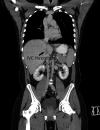
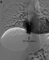
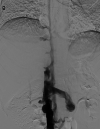
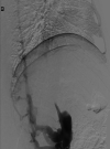
A young adult male was referred for a second opinion of deranged liver biochemistry. He initially presented two years prior with abdominal pain, lethargy and fevers due to a segment two pyogenic liver abscess. He received empirical antibiotic therapy to resolution. Computed tomography for abscess follow-up revealed an intrahepatic inferior vena cava thrombus. He was anti-coagulated with warfarin. He was lupus anticoagulant positive and had a highly positive beta-2 glycoprotein antibody on serial measurement and was diagnosed with anti-phospholipid syndrome. On current review, the patient had no clinical stigmata of chronic liver disease. There were dilated veins on the supraumbilical abdominal and chest walls. There was mild hepatomegaly but no splenomegaly. Laboratory investigations revealed mildly cholestatic liver function tests with hyperbilirubinaemia (40μmol/L) but no liver synthetic dysfunction. Serological screening did not reveal any cause of chronic liver disease. The patient underwent multiphase abdominal CT and formal hepatic venography. What is the diagnosis and describe the hepatic venous outflow?
Keywords: budd chiari syndrome; liver; venous thrombosis.
© Author(s) (or their employer(s)) 2021. Re-use permitted under CC BY-NC. No commercial re-use. See rights and permissions. Published by BMJ.
Conflict of interest statement
Competing interests: None declared.
Figures


References
Publication types
MeSH terms
LinkOut - more resources
Full Text Sources
