. 2022 May 5;59(5):2101293.
doi: 10.1183/13993003.01293-2021. Print 2022 May.

R othia mucilaginosa is an anti-inflammatory bacterium in the respiratory tract of patients with chronic lung disease

Affiliations

R othia mucilaginosa is an anti-inflammatory bacterium in the respiratory tract of patients with chronic lung disease

Charlotte Rigauts et al. Eur Respir J. .

Abstract

Background: Chronic airway inflammation is the main driver of pathogenesis in respiratory diseases such as severe asthma, chronic obstructive pulmonary disease, cystic fibrosis (CF) and bronchiectasis. While the role of common pathogens in airway inflammation is widely recognised, the influence of other microbiota members is still poorly understood.

Methods: We hypothesised that the lung microbiota contains bacteria with immunomodulatory activity which modulate net levels of immune activation by key respiratory pathogens. Therefore, we assessed the immunomodulatory effect of several members of the lung microbiota frequently reported as present in CF lower respiratory tract samples.

Results: We show that Rothia mucilaginosa, a common resident of the oral cavity that is also often detectable in the lower airways in chronic disease, has an inhibitory effect on pathogen- or lipopolysaccharide-induced pro-inflammatory responses, in vitro (three-dimensional cell culture model) and in vivo (mouse model). Furthermore, in a cohort of adults with bronchiectasis, the abundance of Rothia species was negatively correlated with pro-inflammatory markers (interleukin (IL)-8 and IL-1β) and matrix metalloproteinase (MMP)-1, MMP-8 and MMP-9 in sputum. Mechanistic studies revealed that R. mucilaginosa inhibits NF-κB pathway activation by reducing the phosphorylation of IκBα and consequently the expression of NF-κB target genes.

Conclusions: These findings indicate that the presence of R. mucilaginosa in the lower airways potentially mitigates inflammation, which could in turn influence the severity and progression of chronic respiratory disorders.


Conflict of interest statement

Conflict of interest: C. Rigauts declares a patent application related to this work. Conflict of interest: J. Aizawa has nothing to disclose. Conflict of interest: S.L. Taylor has nothing to disclose. Conflict of interest: G.B. Rogers has nothing to disclose. Conflict of interest: M. Govaerts has nothing to disclose. Conflict of interest: P. Cos has nothing to disclose. Conflict of interest: L. Ostyn has nothing to disclose. Conflict of interest: S. Sims has nothing to disclose. Conflict of interest: E. Vandeplassche has nothing to disclose. Conflict of interest: M. Sze has nothing to disclose. Conflict of interest: Y. Dondelinger has nothing to disclose. Conflict of interest: L. Vereecke has nothing to disclose. Conflict of interest: H. Van Acker has nothing to disclose. Conflict of interest: J.L. Simpson has nothing to disclose. Conflict of interest: L. Burr has nothing to disclose. Conflict of interest: A. Willems has nothing to disclose. Conflict of interest: M.M. Tunney has nothing to disclose. Conflict of interest: C. Cigana has nothing to disclose. Conflict of interest: A. Bragonzi has nothing to disclose. Conflict of interest: T. Coenye declares a patent application related to this work. Conflict of interest: A. Crabbé declares a patent application related to this work.

Figures

FIGURE 1
FIGURE 1
Three-dimensional (3D) lung epithelial responses to pro-inflammatory stimuli in the presence and absence of members of the lung microbiota: Pseudomonas aeruginosa PAO1 (Pa), Rothia mucilaginosa DSM20746 (Rm), Staphylococcus aureus SP123 (S), Streptococcus anginosus LMG14696 (St), Achromobacter xylosoxidans LMG26680 (A) and Gemella haemolysans LMG18984 (G). a, b) Interleukin (IL)-8 production by 3D A549 cells after 4 h exposure to a) single bacterial cultures or b) co-cultures of various lung microbiota members with P. aeruginosa PAO1 at an MOI of 30:1. c) IL-8 production by 3D A549 cells after 4 or 24 h exposure to 100 μg·mL−1 lipopolysaccharide (LPS) alone or in co-culture with R. mucilaginosa at an MOI 10:1 (4 h) or 1:1 (24 h). d) IL-6, IL-8, granulocyte–macrophage colony-stimulating factor (GM-CSF) and monocyte chemoattractant protein (MCP)-1 production of 3D A549 cells after 4 h exposure to P. aeruginosa alone or in co-culture with R. mucilaginosa at an MOI of 10:1. NC: negative control (uninfected 3D epithelial cells in serum-free medium). Data represent mean±sem or mean, n≥3. *: p<0.05; **: p<0.001.
FIGURE 2
FIGURE 2
Influence of Rothia mucilaginosa (Rm) on the in vivo responses to lipopolysaccharide (LPS). a) Timeline of animal infection and necropsy, b) macrophage inflammatory protein (MIP)-2 concentration (measured by ELISA), and c) number of CFU·mL−1 in mice lung homogenates after 48 h exposure to vehicle (n=7), LPS (n=18), R. mucilaginosa suspension (n=9 for R. mucilaginosa DSM20746 and n=3 for R. mucilaginosa B03V1S1C) or a combination of LPS+R. mucilaginosa (n=18 for R. mucilaginosa DSM20746 and n=3 for R. mucilaginosa B03V1S1C). d) Lung histopathological score on haematoxylin/eosin-stained sections of the right lung. e) Lung haematoxylin/eosin-stained sections of vehicle, LPS, R. mucilaginosa B03V1S1C and LPS+R. mucilaginosa B03V1S1C instilled mice. Scale bar: 200 μm. Vehicle: sterile alginate beads; LPS: 10 μg per 50 μL. The data presented is from two independent animal experiments for strain DSM20746 and one experiment for strain B03V1S1C. Data represent mean±sem, n≥3. *: p<0.05; **: p<0.01.
FIGURE 3
FIGURE 3
Differential expression of genes involved in inflammation by three-dimensional (3D) A549 alveolar epithelial cells exposed to Pseudomonas aeruginosa versus P. aeruginosa in combination with Rothia mucilaginosa. Quantitative PCR analysis showing average fold changes in mRNA levels of 3D A549 cells stimulated by P. aeruginosa PAO1 versus a co-culture of P. aeruginosa and R. mucilaginosa. n=3. *: statistically significant (p<0.05) fold change.
FIGURE 4
FIGURE 4
Effect of Rothia mucilaginosa (Rm) on NF-κB pathway activation by Pseudomonas aeruginosa (Pa). a) Activation of the NF-κB pathway measured via luminescence of three-dimensional (3D) NF-κB reporter A549 cells. 3D cells were exposed for 4 h to P. aeruginosa PAO1 alone or in co-culture with R. mucilaginosa DSM20746. b) Semiquantitative determination (by Western blotting) of proteins (i.e. A20, IκBα, p65 and phosphorylated (p)-IκBα) produced by 3D A549 cells stimulated with P. aeruginosa PAO1 with or without R. mucilaginosa DSM20746 for 15 min (15′), 30 min (30′), 1 h and 4 h. c) Band intensity (normalised to β-actin) of Western blot at 4 h of 3D A549 cells stimulated with P. aeruginosa PAO1 with or without R. mucilaginosa DSM20746. RLU: relative light units; NC: negative control (uninfected 3D NF-κB reporter A549 cells in serum-free GTSF-2 medium). Data represent mean±sem, n=3. *: p<0.05; **: p<0.01; ***: p<0.001.
FIGURE 5
FIGURE 5
The anti-inflammatory effect of Rothia mucilaginosa (Rm) is mediated by its cell-free supernatant. a) Interleukin (IL)-8 production by three-dimensional (3D) A549 epithelial cells after exposure to P. aeruginosa PAO1 (Pa) for 4 h, with or without R. mucilaginosa DSM20746 live cells, lysed cells or cell-free supernatant. b) Quantification of NF-κB pathway activation after 4 h exposure to lipopolysaccharide (LPS) (100 μg·mL−1) with or without R. mucilaginosa DSM20746 cell-free supernatant. The multiplicity of infection was 10:1 in all experiments. RLU: relative light units; NC: negative control (uninfected 3D epithelial cells). Data represent mean±sem, n≥3. ***: p<0.001.
FIGURE 6
FIGURE 6
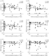
Correlation of absolute load of Rothia mucilaginosa with pro-inflammatory parameters in induced sputum samples from bronchiectasis patients: a) interleukin (IL)-8, b) IL-1β, c) matrix metalloproteinase (MMP)-1, d) MMP-8, e) MMP-9 and f) neutrophils. Data points represent individual induced sputum samples. Correlation coefficients and p-values were calculated on ranked values when Spearman's test was used (rs).

Comment in

References

    1. Tsikrika S, Dimakou K, Papaioannou A, et al. . Non-invasive evaluation of airway inflammation in non-CF bronchiectasis. Eur Respir J 2016; 48: Suppl. 60, PA2556. doi:10.1183/13993003.congress-2016.PA2556 - DOI
    1. Quinn TM, Hill AT. Non-cystic fibrosis bronchiectasis in the elderly: current perspectives. Clin Interv 2018; 13: 1649–1656. doi:10.2147/CIA.S143139 - DOI - PMC - PubMed
    1. Dente FL, Bilotta M, Bartoli ML, et al. . Neutrophilic bronchial inflammation correlates with clinical and functional findings in patients with noncystic fibrosis bronchiectasis. Mediators Inflamm 2015; 2015: 642503. doi:10.1155/2015/642503 - DOI - PMC - PubMed
    1. Murdoch JR, Lloyd CM. Chronic inflammation and asthma. Mutat Res 2010; 690: 24–39. doi:10.1016/j.mrfmmm.2009.09.005 - DOI - PMC - PubMed
    1. Ishmael FT. The inflammatory response in the pathogenesis of asthma. J Am Osteopath Assoc 2011; 111: 11 Suppl. 7, S11–S17. - PubMed

Publication types

LinkOut - more resources