Observational Study
. 2021 Sep 29;11(1):19318.
doi: 10.1038/s41598-021-98201-7.

Proteomic and molecular dynamic investigations of PTM-induced structural fluctuations in breast and ovarian cancer

Affiliations
Observational Study

Proteomic and molecular dynamic investigations of PTM-induced structural fluctuations in breast and ovarian cancer

Dmitry Tikhonov et al. Sci Rep. .

Abstract

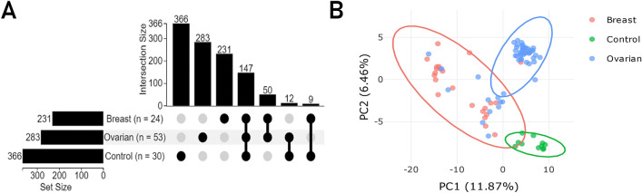
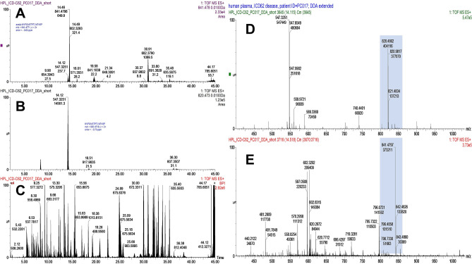
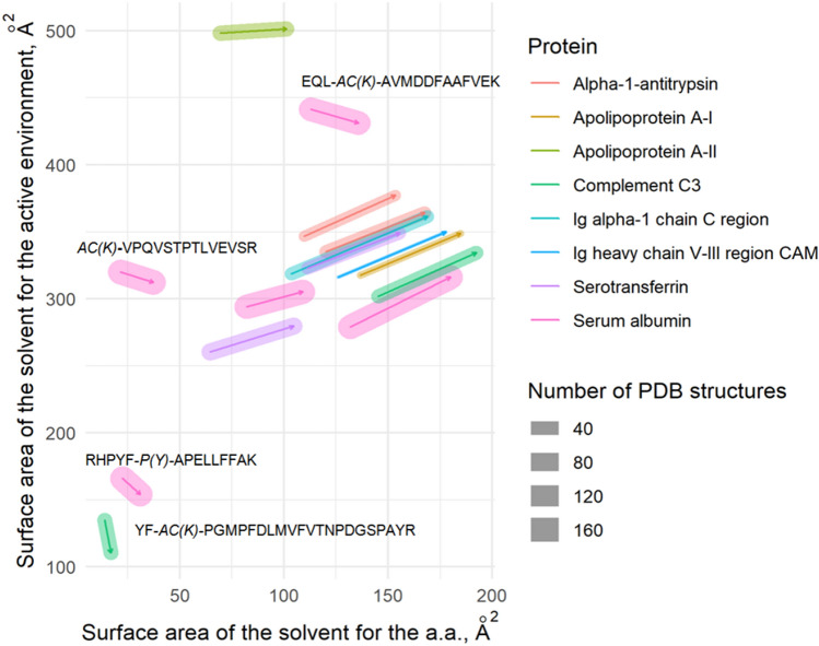
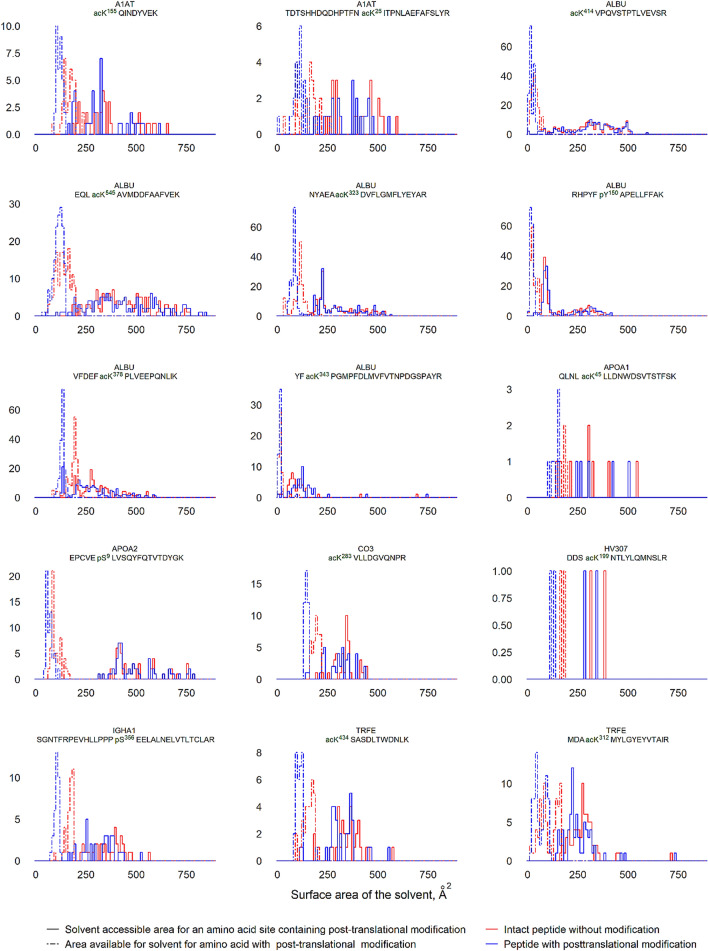
Post-translational processing leads to conformational changes in protein structure that modulate molecular functions and change the signature of metabolic transformations and immune responses. Some post-translational modifications (PTMs), such as phosphorylation and acetylation, are strongly related to oncogenic processes and malignancy. This study investigated a PTM pattern in patients with gender-specific ovarian or breast cancer. Proteomic profiling and analysis of cancer-specific PTM patterns were performed using high-resolution UPLC-MS/MS. Structural analysis, topology, and stability of PTMs associated with sex-specific cancers were analyzed using molecular dynamics modeling. We identified highly specific PTMs, of which 12 modified peptides from eight distinct proteins derived from patients with ovarian cancer and 6 peptides of three proteins favored patients from the group with breast cancer. We found that all defined PTMs were localized in the compact and stable structural motifs exposed outside the solvent environment. PTMs increase the solvent-accessible surface area of the modified moiety and its active environment. The observed conformational fluctuations are still inadequate to activate the structural degradation and enhance protein elimination/clearance; however, it is sufficient for the significant modulation of protein activity.


Conflict of interest statement

The authors declare no competing interests.

Figures

Figure 1
Figure 1
The upset diagram showing the distribution and the intercept size of protein identifications amongst groups of breast and ovarian cancer phenotypes, and the control group (A). PCA analysis carried out for the mutual fraction of proteins (n = 147) with no imputation and reflecting the quantitative alteration of the identified proteins between groups: breast cancer (BC)—red ellipse, ovarian cancer (OC)—blue ellipse and the control (CNT)—green ellipse. The abundancy (quantitative item) was fit to the log-scale (B).
Figure 2
Figure 2
Overview of the targeted mass spectrometry analysis (t-MS2) exemplified on peptide KVPQVSTPTLVEVSR (ALBU) in patient with breast cancer. Extracted ion chromatograms of the modified peptide with m/z = 841.47742+, mass error is -0.83 ppm detected at 14.46 min (A) and its intact (unmodified) counterpart peptide with m/z = 820.47192+, mass error is -0.73 ppm, detected at 14.12 min and shadowed by the unknown peak with m/z = 547.3251 (charge state z = 2 +) (B). Base-peak chromatogram resulted after targeted MS2 analysis shown within analytical time range from 0 to 45 min and 4-folds magnified in intensity scaled (C). Averaged at half of maximum intensity mass spectra of the intact peptide (D) and modified peptide (E). Base peak with m/z = 547.32512+ is visible on the averaged spectra of the intact peptide (D).
Figure 3
Figure 3
Variation of the total solvent-accessible area (in square angstroms; Å2) of active environment after modification of amino acid residue (mounting of PTM). The modified proteins found in plasma samples of patients with breast cancer and ovarian cancer are color-coded. The number of PDB matched structures used for the analysis are designated by bar size.
Figure 4
Figure 4
The distribution of proteins population with the identified PTMs depending on the solvent-accessible area for the certain amino acid residue and its active environment before and after mounting of PTM. The horizontal axis indicated the surface area, accessible for the surrounding solvent (in Å2); the vertical axis indicated the number of the affected protein molecules. Color-code defines amino acids before modification (blue dashed line), amino acids after modification (pink dashed line), active environment before modification (blue solid line) and active environment after modification (pink solid line).
Figure 5
Figure 5
Calculated geometrical features of albumin (ALBU) motives with PTM moiety. Each motif is characterized by the estimated geometrical features for intact molecule (stat; blue color), calculated results of molecular dynamics for the unmodified molecule (nmd; green color), and calculated features for the modified molecule (mod; red color). The right axis indicates (d) distances between helices, (r) inter-planar distance between helices, S—area of the spatial projection, and θ—torsion angle.

References

    1. Breast Cancer: Symptoms and Causes. Mayo Clinic https://www.mayoclinic.org/diseases-conditions/breast-cancer/symptoms-ca... (accessed 25 Jan 2021).
    1. Breast Cancer Statistics | World Cancer Research Fund https://www.wcrf.org/dietandcancer/cancer-trends/breast-cancer-statistics (accessed 25 Jan 2021).
    1. Sharma BS, Prabhakaran V, Desai AP, Bajpai J, Verma RJ, Swain PK. Post-translational modifications (PTMs), from a cancer perspective: An overview. Oncogen. 2019 doi: 10.35702/onc.10012. - DOI
    1. Venne AS, Kollipara L, Zahedi RP. The next level of complexity: Crosstalk of posttranslational modifications. Proteomics. 2014;14(4–5):513–524. doi: 10.1002/pmic.201300344. - DOI - PubMed
    1. Brentville VA, Vankemmelbeke M, Metheringham RL, Durrant LG. Post-Translational modifications such as citrullination are excellent targets for cancer therapy. Semin. Immunol. 2020;47:101393. doi: 10.1016/j.smim.2020.101393. - DOI - PubMed

Publication types

MeSH terms