Review
. 2021 Sep 7;27(33):5460-5473.
doi: 10.3748/wjg.v27.i33.5460.

Guidelines on postoperative magnetic resonance imaging in patients operated for cryptoglandular anal fistula: Experience from 2404 scans

Affiliations
Review

Guidelines on postoperative magnetic resonance imaging in patients operated for cryptoglandular anal fistula: Experience from 2404 scans

Pankaj Garg et al. World J Gastroenterol. .

Abstract

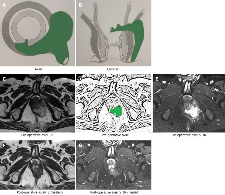
Magnetic resonance imaging (MRI) is considered the gold standard for the evaluation of anal fistulas. There is sufficient literature available outlining the interpretation of fistula MRI before performing surgery. However, the interpretation of MRI becomes quite challenging in the postoperative period after the surgery of fistula has been undertaken. Incidentally, there are scarce data and no set guidelines regarding analysis of fistula MRI in the postoperative period. In this article, we discuss the challenges faced while interpreting the postoperative MRI, the timing of the postoperative MRI, the utility of MRI in the postoperative period for the management of anal fistulas, the importance of the active involvement and experience of the treating clinician in interpreting MRI scans, and the latest advancements in the field.

Keywords: Anal fistula; Hyperintensity; Internal opening; Magnetic resonance imaging; Postoperative.


Conflict of interest statement

Conflict-of-interest statement: None of the authors, Garg P, Kaur B, Yagnik VD, Dawka S and Menon GR, have any conflict of interest.

Figures

Figure 1
Figure 1
A 37-year-old male patient with a high posterior intersphincteric abscess was drained by transanal opening of the intersphincteric space procedure. Magnetic resonance imaging (MRI) done after 3 wk after surgery showed hyperintense signal (due to healing granulation tissue) at the same location similar to the hyperintense signal in the preoperative images (due to pus collection). A: Axial section (schematic diagram); B: Coronal section (schematic diagram); C: Pre-operative T2-weighted MRI; D: Sketch of pre-operative axial MRI image highlighting abscess (green color); E: Pre-operative short-T1 inversion recovery (STIR) axial MRI; F: Pre-operative T2-weighted coronal MRI; G: Sketch of pre-operative coronal MRI image highlighting abscess (green color); H: Postoperative T2-weighted axial MRI; I: Sketch of postoperative axial MRI image highlighting healing granulation tissue (green color); J: Postoperative STIR axial MRI; K: Postoperative T2-weighted coronal MRI; L: Sketch of postoperative coronal MRI image highlighting healing granulation tissue (green color). STIR: Short-T1 inversion recovery.
Figure 2
Figure 2
A 64-year-old male patient with a high transsphincteric fistula with multiple branches. Magnetic resonance imaging (MRI) done after 19 wk post-surgery showing fistula has healed completely. The hyperintense signal (white) in the preoperative images has turned hypointense (black). A: Axial section (schematic diagram); B: Coronal section (schematic diagram); C: Pre-operative T2-weighted axial MRI; D: Sketch of pre-operative axial MRI image highlighting internal opening and the intersphincteric portion of fistula tract (green color); E: Pre-operative short-T1 inversion recovery (STIR) axial MRI; F: Postoperative T2-weighted axial MRI (after 19 wk) showing completely healed fistula; G: Postoperative STIR axial MRI (after 19 wk) showing completely healed fistula. STIR: Short-T1 inversion recovery.
Figure 3
Figure 3
A 54-year-old male patient with a high transsphincteric fistula. The fistula appeared to have ‘clinically’ healed as the external opening had closed with cessation of all pus. However, magnetic resonance imaging (MRI) done after 28 wk post-surgery showed patent internal opening and widening of intersphincteric portion of fistula tract. A: Axial section (schematic diagram); B: Coronal section (schematic diagram); C: Pre-operative T2-weighted MRI; D: Sketch of pre-operative axial MRI image highlighting internal opening and the intersphincteric portion of fistula tract (green color); E: Pre-operative short-T1 inversion recovery (STIR) axial MRI; F: Postoperative T2-weighted axial MRI (after 28 wk) showing patent internal opening and persistent intersphincteric portion of fistula tract; G: Sketch of postoperative axial MRI image highlighting patent internal opening and persistent intersphincteric portion of fistula tract; H: Postoperative STIR axial MRI (after 28 wk) showing patent internal opening and persistent intersphincteric portion of fistula tract. STIR: Short-T1 inversion recovery.
Figure 4
Figure 4
A 47-year-old male patient with a high transsphincteric fistula with supralevator extension. The fistula appeared to have ‘clinically’ healed as the external opening had closed with cessation of all pus. Magnetic resonance imaging (MRI) done after 26 wk post-surgery showed patent internal opening and active intersphincteric portion of the fistula tract [hyperintense on T2 and short-T1 inversion recovery (STIR)]. The patient was operated again. MRI done after 14 wk after 2nd surgery showed fistula has healed completely. The hyperintense signal (white) in the preoperative images has turned hypointense (black). A: Axial section (schematic diagram); B: Coronal section (schematic diagram); C: Pre-operative T2-weighted MRI; D: Sketch of pre-operative axial MRI image highlighting internal opening and the intersphincteric portion of fistula tract (green color); E: Pre-operative STIR axial MRI; F: Postoperative (26 wk after 1st surgery) T2-weighted axial MRI showing patent internal opening and persistent intersphincteric portion of fistula tract; G: Sketch highlighting patent internal opening and intersphincteric portion of fistula tract (green color); H: Postoperative (26 wk after 1st surgery) STIR axial MRI showing patent internal opening and persistent intersphincteric portion of fistula tract; I: Postoperative (14 wk after 2nd surgery) T2-weighted axial MRI showing completely healed fistula; J: Postoperative (14 wk after 2nd surgery) STIR axial MRI showing completely healed fistula. STIR: Short-T1 inversion recovery.
Figure 5
Figure 5
A 51-year-old male patient with a high transsphincteric fistula with multiple branches. Magnetic resonance imaging (MRI) done after 8 wk post-surgery showing fistula has healed completely. The hyperintense signal (white) in the preoperative images has turned hypointense (black). A: Axial section (schematic diagram); B: Coronal section (schematic diagram); C: Pre-operative T2-weighted axial MRI; D: Sketch of pre-operative axial MRI image highlighting internal opening, the intersphincteric portion of fistula tract and transsphincteric fistula tract (green color); E: Pre-operative short-T1 inversion recovery (STIR) axial MRI; F: Postoperative T2-weighted axial MRI (after 8 wk) showing completely healed fistula; G: Postoperative STIR axial MRI (after 8 wk) showing completely healed fistula. STIR: Short-T1 inversion recovery.
Figure 6
Figure 6
A 40-year-old male patient with a suprasphincteric fistula. Magnetic resonance imaging (MRI) done after 16 wk post-surgery showing fistula has healed completely. The hyperintense signal (white) in the preoperative images has turned hypointense (black). A: Axial section (schematic diagram); B: Coronal section (schematic diagram); C: Pre-operative T2-weighted axial MRI; D: Sketch of pre-operative axial MRI image highlighting internal opening and the intersphincteric portion of fistula tract (green color); E: Pre-operative short-T1 inversion recovery (STIR) axial MRI; F: Postoperative T2-weighted axial MRI (after 16 wk) showing completely healed fistula; G: Postoperative STIR axial MRI (after 16 wk) showing completely healed fistula. STIR: Short-T1 inversion recovery.
Figure 7
Figure 7
A 69-year-old male patient with a posterior fistula with a presacral abscess. A wide bore drainage tube was inserted into the cavity intraoperatively. However, the patient did not improve. Postoperative check magnetic resonance imaging (MRI) demonstrated that the tube was in the wrong plane and the presacral abscess was not properly drained. A: Axial section (schematic diagram); B: Coronal section (schematic diagram); C: Preoperative short-T1 inversion recovery (STIR) sagittal MRI; D: Sketch of pre-operative sagittal MRI image highlighting anal fistula and presacral abscess (green color); E: Postoperative STIR sagittal MRI showing drainage tube in the wrong plane and persistent presacral abscess; F: Sketch of postoperative sagittal MRI highlighting persisting anal fistula and presacral abscess (green color) (orange arrows show the drainage tube). STIR: Short-T1 inversion recovery.

References

    1. Włodarczyk M, Włodarczyk J, Sobolewska-Włodarczyk A, Trzciński R, Dziki Ł, Fichna J. Current concepts in the pathogenesis of cryptoglandular perianal fistula. J Int Med Res. 2021;49:300060520986669. - PMC - PubMed
    1. Mei Z, Li Y, Zhang Z, Zhou H, Liu S, Han Y, Du P, Qin X, Shao Z, Ge M, Wang Q, Yang W. Development of screening tools to predict the risk of recurrence and related complications following anal fistula surgery: protocol for a prospective cohort study. BMJ Open. 2020;10:e035134. - PMC - PubMed
    1. Mei Z, Wang Q, Zhang Y, Liu P, Ge M, Du P, Yang W, He Y. Risk Factors for Recurrence after anal fistula surgery: A meta-analysis. Int J Surg. 2019;69:153–164. - PubMed
    1. Terra MP, Beets-Tan RG, van der Hulst VP, Deutekom M, Dijkgraaf MG, Bossuyt PM, Dobben AC, Baeten CG, Stoker J. MRI in evaluating atrophy of the external anal sphincter in patients with fecal incontinence. AJR Am J Roentgenol. 2006;187:991–999. - PubMed
    1. Garcia-Aguilar J, Belmonte C, Wong WD, Goldberg SM, Madoff RD. Anal fistula surgery. Factors associated with recurrence and incontinence. Dis Colon Rectum. 1996;39:723–729. - PubMed